Blood pressure
From Wikipedia, the free encyclopedia
- "Blood Pressure" is also the title of a short story by Damon Runyan in "Guys and Dolls and Other Stories".
- See Hypertension for more information about high blood pressure.

Blood pressure (BP) is the pressure (force per unit area) exerted by circulating blood on the walls of blood vessels, and constitutes one of the principal vital signs. The pressure of the circulating blood decreases as it moves away from the heart through arteries and capillaries, and toward the heart through veins. When unqualified, the term blood pressure usually refers to brachial arterial pressure: that is, in the major blood vessel of the upper left or right arm that takes blood away from the heart. Blood pressure may, however, sometimes be measured at other sites in the body, for instance at the ankle. The ratio of the blood pressure measured in the main artery at the ankle to the brachial blood pressure gives the Ankle Brachial Pressure Index (ABPI).
Contents |
[edit] Measurement
Arterial pressure is most commonly measured via a sphygmomanometer, which historically used the height of a column of mercury to reflect the circulating pressure (see Noninvasive measurement). Today blood pressure values are still reported in millimetres of mercury (mmHg), though aneroid and electronic devices do not use mercury.
For each heartbeat, blood pressure varies between systolic and diastolic pressures. Systolic pressure is peak pressure in the arteries, which occurs near the beginning of the cardiac cycle when the ventricles are contracting. Diastolic pressure is minimum pressure in the arteries, which occurs near the end of the cardiac cycle when the ventricles are filled with blood. An example of normal measured values for a resting, healthy adult human is 115 mmHg systolic and 75 mmHg diastolic (written as 115/75 mmHg, and spoken (in the US) as "one fifteen over seventy-five"). Pulse pressure is the difference between systolic and diastolic pressures.
Systolic and diastolic arterial blood pressures are not static but undergo natural variations from one heartbeat to another and throughout the day (in a circadian rhythm). They also change in response to stress, nutritional factors, drugs, disease, exercise, and momentarily from standing up. Sometimes the variations are large. Hypertension refers to arterial pressure being abnormally high, as opposed to hypotension, when it is abnormally low. Along with body temperature, blood pressure measurements are the most commonly measured physiological parameters.
Arterial pressures can be measured invasively (by penetrating the skin and measuring inside the blood vessels) or non-invasively. The former is usually restricted to a hospital setting.
[edit] Units
The predominantly used unit for blood pressure measurement is mmHg (millimeter of mercury). For example, normal pressure can be stated as 120 over 80.
[edit] Noninvasive measurement
The noninvasive auscultatory (from the Latin for listening) and oscillometric measurements are simpler and quicker than invasive measurements, require less expertise in fitting, have virtually no complications, and are less unpleasant and painful for the patient. However, non-invasive measures may yield somewhat lower accuracy and small systematic differences in numerical results. Non-invasive measurement methods are more commonly used for routine examinations and monitoring.
[edit] Palpation methods
A minimum systolic value can be roughly estimated without any equipment by palpation, most often used in emergency situations. Palpation of a radial pulse indicates a minimum blood pressure of 80 mmHg, a femoral pulse indicates at least 70 mmHg, and a carotid pulse indicates a minimum of 60 mmHg. However, one study indicated that this method was not accurate enough and often overestimated patients' systolic blood pressure.[1] A more accurate value of systolic blood pressure can be obtained with a sphygmomanometer and palpating for when a radial pulse returns.[2] The diastolic blood pressure can not be estimated by this method.[3]
[edit] Auscultatory methods

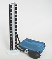
The auscultatory method uses a stethoscope and a sphygmomanometer. This comprises an inflatable (Riva-Rocci) cuff placed around the upper arm at roughly the same vertical height as the heart, attached to a mercury or aneroid manometer. The mercury manometer, considered to be the gold standard for arterial pressure measurement[citation needed], measures the height of a column of mercury, giving an absolute result without need for calibration, and consequently not subject to the errors and drift of calibration which affect other methods. The use of mercury manometers is often required in clinical trials and for the clinical measurement of hypertension in high risk patients, such as pregnant women.
A cuff of appropriate size is fitted smoothly and snugly,then inflated manually by repeatedly squeezing a rubber bulb until the artery is completely occluded. Listening with the stethoscope to the brachial artery at the elbow, the examiner slowly releases the pressure in the cuff. When blood just starts to flow in the artery, the turbulent flow creates a "whooshing" or pounding (first Korotkoff sound). The pressure at which this sound is first heard is the systolic blood pressure. The cuff pressure is further released until no sound can be heard (fifth Korotkoff sound), at the diastolic arterial pressure. Sometimes, the pressure is palpated (felt by hand) to get an estimate before auscultation.
[edit] Oscillometric methods
Oscillometric methods are sometimes used in the long-term measurement and sometimes in general practice. The equipment is functionally similar to that of the auscultatory method, but with an electronic pressure sensor (transducer) fitted in to detect blood flow, instead of using the stethoscope and the expert's ear. In practice, the pressure sensor is a calibrated electronic device with a numerical readout of blood pressure. To maintain accuracy, calibration must be checked periodically, unlike the inherently accurate mercury manometer. In most cases the cuff is inflated and released by an electrically operated pump and valve, which may be fitted on the wrist (elevated to heart height), although the upper arm is preferred. They vary widely in accuracy, and should be checked at specified intervals and if necessary recalibrated.
Oscillometric measurement requires less skill than the auscultatory technique, and may be suitable for use by untrained staff and for automated patient home monitoring.
The cuff is inflated to a pressure initially in excess of the systolic arterial pressure, and then reduces to below diastolic pressure over a period of about 30 seconds. When blood flow is nil (cuff pressure exceeding systolic pressure) or unimpeded (cuff pressure below diastolic pressure), cuff pressure will be essentially constant. It is essential that the cuff size is correct: undersized cuffs may yield too high a pressure, whereas oversized cuffs yield too low a pressure. When blood flow is present, but restricted, the cuff pressure, which is monitored by the pressure sensor, will vary periodically in synchrony with the cyclic expansion and contraction of the brachial artery, i.e., it will oscillate. The values of systolic and diastolic pressure are computed, not actually measured from the raw data, using an algorithm; the computed results are displayed.
Oscillometric monitors may produce inaccurate readings in patients with heart and circulation problems, that include arterial sclerosis, arrhythmia, preeclampsia, pulsus alternans, and pulsus paradoxus.
In practice the different methods do not give identical results; an algorithm and experimentally obtained coefficients are used to adjust the oscillometric results to give readings which match the auscultatory results as well as possible. Some equipment uses computer-aided analysis of the instantaneous arterial pressure waveform to determine the systolic, mean, and diastolic points. Since many oscillometric devices have not been validated, caution must be given as most are not suitable in clinical and acute care settings.
The term NIBP, for Non-Invasive Blood Pressure, is often used to describe oscillometric monitoring equipment.
[edit] Invasive measurement
Arterial blood pressure (BP) is most accurately measured invasively through an arterial line. Invasive arterial pressure measurement with intravascular cannulae involves direct measurement of arterial pressure by placing a cannula needle in an artery (usually radial, femoral, dorsalis pedis or brachial). This procedure can be done by any licensed MD or an RT (Respiratory Therapist).
The cannula must be connected to a sterile, fluid-filled system, which is connected to an electronic pressure transducer. The advantage of this system is that pressure is constantly monitored beat-by-beat, and a waveform (a graph of pressure against time) can be displayed. This invasive technique is regularly employed in human and veterinary intensive care medicine, anesthesiology, and for research purposes.
Cannulation for invasive vascular pressure monitoring is infrequently associated with complications such as thrombosis, infection, and bleeding. Patients with invasive arterial monitoring require very close supervision, as there is a danger of severe bleeding if the line becomes disconnected. It is generally reserved for patients where rapid variations in arterial pressure are anticipated.
Invasive vascular pressure monitors are pressure monitoring systems designed to acquire pressure information for display and processing. There are a variety of invasive vascular pressure monitors for trauma, critical care, and operating room applications. These include single pressure, dual pressure, and multi-parameter (i.e. pressure / temperature). The monitors can be used for measurement and follow-up of arterial, central venous, pulmonary arterial, left atrial, right atrial, femoral arterial, umbilical venous, umbilical arterial, and intracranial pressures.
Vascular pressure parameters are derived in the monitor's microcomputer system. Usually, systolic, diastolic, and mean pressures are displayed simultaneously for pulsatile waveforms (i.e. arterial and pulmonary arterial). Some monitors also calculate and display CPP (cerebral perfusion pressure). Normally, a zero key on the front of the monitor makes pressure zeroing extremely fast and easy. Alarm limits may be set to assist the medical professional responsible for observing the patient. High and low alarms may be set on displayed temperature parameters.
[edit] Home monitoring
For some patients, blood pressure measurements taken in a doctor's office may not correctly characterize their typical blood pressure. In up to 25% of patients, the office visit blood pressure reading is higher than their typical blood pressure. This type of error is called white coat hypertension and can result from anxiety related to an examination by a health care professional.[4] The misdiagnosis of hypertension for these patients can result in needless and possibly harmful medication. On the other hand, in some cases a lower than typical blood pressure reading occurs at the doctor's office and these patients may fail to get needed treatment for hypertension.[5]
Ambulatory blood pressure devices that take readings every half hour throughout the day and night have been used for identifying and mitigating these problems. Except for periods during sleep, home monitoring could be used for these purposes instead of ambulatory blood pressure monitoring.[6] Home monitoring may also be used to improve hypertension management and to monitor the effects of lifestyle changes and medication related to blood pressure.[7] Compared to ambulatory blood pressure measurements, home monitoring has been found to be an effective and lower cost alternative.[6][8][9]
Aside from the white coat effect, arterial pressure readings outside of a clinical setting are usually slightly lower in the majority of people. The studies that looked into the risks from hypertension and the benefits of lowering the arterial pressure in affected patients were based on readings in a clinical environment.
When measuring blood pressure, an accurate reading requires that one not drink coffee, smoke cigarettes, or engage in strenuous exercise for 30 minutes before taking the reading. A full bladder may have a small effect on blood pressure readings, so if the urge to urinate exists, one should do so before the reading. For 5 minutes before the reading, one should sit upright in a chair with one's feet flat on the floor and with limbs uncrossed. The blood pressure cuff should always be against bare skin, as readings taken over a shirt sleeve are less accurate. During the reading, the arm that is used should be relaxed and kept at heart level, for example by resting it on a table.[10]
Since arterial pressure varies throughout the day, measurements intended to monitor changes over longer time frames should be taken at the same time of day to ensure that the readings are comparable. Suitable times are:
- immediately after awakening (before washing/dressing and taking breakfast/drink), while the body is still resting,
- immediately after finishing work.
Automatic self-contained blood pressure monitors are available at reasonable prices, some of which are capable of Korotkoff's measurement in addition to oscillometric methods, enabling irregular heartbeat patients to accurately measure their blood pressure at home.
[edit] Classification
The following classification of blood pressure applies to adults aged 18 and older. It is based on the average of seated blood pressure readings that were properly measured during 2 or more office visits.[7][11]
| Category | systolic, mmHg | diastolic, mmHg |
|---|---|---|
|
|
|
|
|
|
|
|
|
|
|
|
|
|
|
|
|
|
|
|
[edit] Normal values
While average values for arterial pressure could be computed for any given population, there is often a large variation from person to person; arterial pressure also varies in individuals from moment to moment. Additionally, the average of any given population may have a questionable correlation with its general health, thus the relevance of such average values is equally questionable. However, in a study of 100 subjects with no known history of hypertension, an average blood pressure of 112/64 mmHg was found,[12] which is in the normal range.
In children the normal ranges are lower than for adults.[13] In the elderly, blood pressure tends to be higher than normal adult values, largely because of reduced flexibility of the arteries. Factors such as age and gender[14] influence blood pressure values. Pressure also varies with exercise, emotional reactions, sleep, digestion and time of day.
Differences between left and right arm blood pressure measurements tend to be random and average to nearly zero if enough measurements are taken. However, in a small percentage of cases there is a consistently present difference greater than 10 mmHg which may need further investigation, e.g. for obstructive arterial disease.[15][16]
The risk of cardiovascular disease increases progressively throughout the range of higher arterial pressure that begins at 115/75 mmHg.[17] In the past, hypertension was only diagnosed if secondary signs of high arterial pressure were present, along with a prolonged high systolic pressure reading over several visits. In the US, this reactive stance has been soundly rejected in light of recent evidence[citation needed]. However in the UK, patients’ readings are still considered normal up to 140/90 mmHg.[18]
Clinical trials demonstrate that people who maintain arterial pressures at the low end of these pressure ranges have much better long term cardiovascular health. The principal medical debate concerns the aggressiveness and relative value of methods used to lower pressures into this range for those who do not maintain such pressure on their own. Elevations, more commonly seen in older people, though often considered normal, are associated with increased morbidity and mortality. The clear trend from double blind clinical trials (for the better strategies and agents) demonstrates that lower arterial pressure correlates with lower rates of disease.[citation needed]
[edit] Physiology
The physics of the circulatory system are very complex. That said, there are many physical factors that influence arterial pressure. Each of these may in turn be influenced by physiological factors, such as diet, exercise, disease, drugs or alcohol, obesity, excess weight and so-forth.
Some physical factors are:
- Rate of pumping. In the circulatory system, this rate is called heart rate, the rate at which blood (the fluid) is pumped by the heart. The volume of blood flow from the heart is called the cardiac output which is the heart rate (the rate of contraction) multiplied by the stroke volume (the amount of blood pumped out from the heart with each contraction). The higher the heart rate, the higher the arterial pressure, assuming no reduction in stroke volume.
- Volume of fluid or blood volume, the amount of blood that is present in the body. The more blood present in the body, the higher the rate of blood return to the heart and the resulting cardiac output. There is some relationship between dietary salt intake and increased blood volume, potentially resulting in higher arterial pressure, though this varies with the individual and is highly dependent on autonomic nervous system response and the renin-angiotensin system.
- Resistance. In the circulatory system, this is the resistance of the blood vessels. The higher the resistance, the higher the arterial pressure upstream from the resistance to blood flow. Resistance is related to vessel radius (the larger the radius, the lower the resistance), vessel length (the longer the vessel, the higher the resistance), as well as the smoothness of the blood vessel walls. Smoothness is reduced by the build up of fatty deposits on the arterial walls. Substances called vasoconstrictors can reduce the size of blood vessels, thereby increasing blood pressure. Vasodilators (such as nitroglycerin) increase the size of blood vessels, thereby decreasing arterial pressure. Resistance, and its relation to volumetric flow rate (Q) and pressure difference between the two ends of a vessel are described by Poiseuille's Law.
- Viscosity, or thickness of the fluid. If the blood gets thicker, the result is an increase in arterial pressure. Certain medical conditions can change the viscosity of the blood. For instance, low red blood cell concentration, anemia, reduces viscosity, whereas increased red blood cell concentration increases viscosity. Viscosity also increases with blood sugar concentration—visualize pumping syrup. It had been thought that aspirin and related "blood thinner" drugs decreased the viscosity of blood, but studies found[19] that they act by reducing the tendency of the blood to clot instead.
In practice, each individual's autonomic nervous system responds to and regulates all these interacting factors so that, although the above issues are important, the actual arterial pressure response of a given individual varies widely because of both split-second and slow-moving responses of the nervous system and end organs. These responses are very effective in changing the variables and resulting blood pressure from moment to moment.
[edit] Mean arterial pressure
The mean arterial pressure (MAP) is the average over a cardiac cycle and is determined by the cardiac output (CO), systemic vascular resistance (SVR), and central venous pressure (CVP),[20]

MAP can be approximately determined from measurements of the systolic pressure  and the diastolic pressure
  while there is a normal resting heart rate,[20]

[edit] Pulse pressure
The up and down fluctuation of the arterial pressure results from the pulsatile nature of the cardiac output, i.e. the heartbeat. The pulse pressure is determined by the interaction of the stroke volume of the heart, compliance (ability to expand) of the aorta, and the resistance to flow in the arterial tree. By expanding under pressure, the aorta absorbs some of the force of the blood surge from the heart during a heartbeat. In this way the pulse pressure is reduced from what it would be if the aorta wasn't compliant.[21]
The pulse pressure can be simply calculated from the difference of the measured systolic and diastolic pressures,[21]

[edit] Vascular resistance
The larger arteries, including all large enough to see without magnification, are low resistance conduits (assuming no advanced atherosclerotic changes) with high flow rates that generate only small drops in pressure. For instance, with a subject in the supine position, blood traveling from the heart to the toes typically only experiences a 5 mmHg drop in mean pressure[citation needed].
[edit] Vascular pressure wave
Modern physiology developed the concept of the vascular pressure wave (VPW). This wave is created by the heart during the systole and originates in the ascending aorta. Much faster than the stream of blood itself, it is then transported through the vessel walls to the peripheral arteries. There the pressure wave can be palpated as the peripheral pulse. As the wave is reflected at the peripheral veins it runs back in a centripetal fashion. Where the crests of the reflected and the original wave meet, the pressure inside the vessel is higher than the true pressure in the aorta. This concept explains why the arterial pressure inside the peripheral arteries of the legs and arms is higher than the arterial pressure in the aorta,[22][23][24] and in turn for the higher pressures seen at the ankle compared to the arm with normal ankle brachial pressure index values.
[edit] Regulation
The endogenous regulation of arterial pressure is not completely understood. Currently, three mechanisms of regulating arterial pressure have been well-characterized:
- Baroreceptor reflex: Baroreceptors detect changes in arterial pressure and send signals ultimately to the medulla of the brain stem. The medulla, by way of the autonomic nervous system, adjusts the mean arterial pressure by altering both the force and speed of the heart's contractions, as well as the total peripheral resistance. The most important arterial baroreceptors are located in the left and right carotid sinuses and in the aortic arch.[25]
- Renin-angiotensin system (RAS): This system is generally known for its long-term adjustment of arterial pressure. This system allows the kidney to compensate for loss in blood volume or drops in arterial pressure by activating an endogenous vasoconstrictor known as angiotensin II.
- Aldosterone release: This steroid hormone is released from the adrenal cortex in response to angiotensin II or high serum potassium levels. Aldosterone stimulates sodium retention and potassium excretion by the kidneys. Since sodium is the main ion that determines the amount of fluid in the blood vessels by osmosis, aldosterone will increase fluid retention, and indirectly, arterial pressure.
These different mechanisms are not necessarily independent of each other, as indicated by the link between the RAS and aldosterone release. Currently, the RAS system is targeted pharmacologically by ACE inhibitors and angiotensin II receptor antagonists. The aldosterone system is directly targeted by spironolactone, an aldosterone antagonist. The fluid retention may be targeted by diuretics; the antihypertensive effect of diuretics is due to its effect on blood volume. Generally, the baroreceptor reflex is not targeted in hypertension because if blocked, individuals may suffer from orthostatic hypotension and fainting.
[edit] Pathophysiology
[edit] High arterial pressure

Arterial hypertension can be an indicator of other problems and may have long-term adverse effects. Sometimes it can be an acute problem, for example hypertensive emergency.
All levels of arterial pressure put mechanical stress on the arterial walls. Higher pressures increase heart workload and progression of unhealthy tissue growth (atheroma) that develops within the walls of arteries. The higher the pressure, the more stress that is present and the more atheroma tend to progress and the heart muscle tends to thicken, enlarge and become weaker over time.
Persistent hypertension is one of the risk factors for strokes, heart attacks, heart failure and arterial aneurysms, and is the leading cause of chronic renal failure. Even moderate elevation of arterial pressure leads to shortened life expectancy. At severely high pressures, mean arterial pressures 50% or more above average, a person can expect to live no more than a few years unless appropriately treated.[26]
In the past, most attention was paid to diastolic pressure; but nowadays it is recognised that both high systolic pressure and high pulse pressure (the numerical difference between systolic and diastolic pressures) are also risk factors. In some cases, it appears that a decrease in excessive diastolic pressure can actually increase risk, due probably to the increased difference between systolic and diastolic pressures (see the article on pulse pressure).
[edit] Low arterial pressure
Blood pressure that is too low is known as hypotension. The similarity in pronunciation with hypertension can cause confusion. Hypotension is a medical concern only if it causes signs or symptoms, such as dizziness, fainting, or in extreme cases, shock.[11]
When arterial pressure and blood flow decrease beyond a certain point, the perfusion of the brain becomes critically decreased (i.e., the blood supply is not sufficient), causing lightheadedness, dizziness, weakness or fainting.
Sometimes the arterial pressure drops significantly when a patient stands up from sitting. This is known as orthostatic hypotension (postural hypotension); gravity reduces the rate of blood return from the body veins below the heart back to the heart, thus reducing stroke volume and cardiac output.
When people are healthy, the veins below their heart quickly constrict and the heart rate increases to minimize and compensate for the gravity effect. This is carried out involuntarily by the autonomic nervous system. The system usually requires a few seconds to fully adjust and if the compensations are too slow or inadequate, the individual will suffer reduced blood flow to the brain, dizziness and potential blackout. Increases in G-loading, such as routinely experienced by acrobatic jet pilots 'pulling Gs', greatly increases this effect. Repositioning the body perpendicular to gravity largely eliminates the problem.
Other causes of low arterial pressure include:
- Sepsis
- Hemorrhage - blood loss
- Toxins including toxic doses of blood pressure medicine
- Hormonal abnormalities, such as Addison's disease
Shock is a complex condition which leads to critically decreased perfusion. The usual mechanisms are loss of blood volume, pooling of blood within the veins reducing adequate return to the heart and/or low effective heart pumping. Low arterial pressure, especially low pulse pressure, is a sign of shock and contributes to and reflects decreased perfusion.
If there is a significant difference in the pressure from one arm to the other, that may indicate a narrowing (for example, due to aortic coarctation, aortic dissection, thrombosis or embolism) of an artery.
[edit] Venous pressure
Venous pressure is the vascular pressure in a vein or in the atria of the heart. It is much less than arterial pressure, with common values of 5 mmHg in the right atrium and 8 mmHg in the left atrium. Measurement of pressures in the venous system and the pulmonary vessels plays an important role in intensive care medicine but requires an invasive central venous catheter.
[edit] See also
[edit] Footnotes
- ^ Deakin CD, Low JL (September 2000). "Accuracy of the advanced trauma life support guidelines for predicting systolic blood pressure using carotid, femoral, and radial pulses: observational study". BMJ 321 (7262): 673–4. PMID 10987771. PMC: 27481. http://bmj.com/cgi/pmidlookup?view=long&pmid=10987771.
- ^ Interpretation - Blood Pressure - Vitals, "University of Florida". Retrieved on 2008-03-18.
- ^ G8 Secondary Survey, "Manitoba". Retrieved on 2008-03-18.
- ^ Jhalani, Juhee a; Goyal, Tanya a; Clemow, Lynn a; Schwartz, Joseph E. b; Pickering, Thomas G. a; Gerin, William a. Anxiety and outcome expectations predict the white-coat effect.. 10(6), December 2005. Lippincott Williams & Wilkins, Inc.. pp. pp317-319. http://www.bpmonitoring.com/pt/re/bpm/abstract.00126097-200512000-00006.htm;jsessionid=LpvGzJN7PDC1yqJtnQj3ZWfmzgdnhWycyzsKybSHsr2FLx3hR1vh!1805002056!181195629!8091!-1.
- ^ Elliot, Victoria Stagg (2007-06-11). "Blood pressure readings often unreliable". American Medical News (American Medical Association). http://www.ama-assn.org/amednews/2007/06/11/hlsa0611.htm. Retrieved on 2008-08-16.
- ^ a b Mancia G, De Backer G, Dominiczak A, et al (June 2007). "2007 Guidelines for the management of arterial hypertension: The Task Force for the Management of Arterial Hypertension of the European Society of Hypertension (ESH) and of the European Society of Cardiology (ESC)". Eur Heart J 28 (12): 1462–536. doi:. PMID 17562668.
- ^ a b Chobanian AV, Bakris GL, Black HR, et al (December 2003). "Seventh report of the Joint National Committee on Prevention, Detection, Evaluation, and Treatment of High Blood Pressure". Hypertension 42 (6): 1206–52. doi:. PMID 14656957.
- ^ Niiranen, TJ; Kantola IM, Vesalainen R, et al (2006). "A comparison of home measurement and ambulatory monitoring of blood pressure in the adjustment of antihypertensive treatment". Am J Hypertens 19 (5): 468–74. doi:. PMID 16647616.
- ^ Shimbo, Daichi; Thomas G. Pickering, Tanya M. Spruill, et al (2007). "Relative utility of home, ambulatory, and office blood pressures in the prediction of end-organ damage". Am J Hypertens 20 (5): 476–82. doi:. PMID 17485006. http://www.nature.com/ajh/journal/v20/n5/abs/ajh200783a.html.
- ^ National Heart, Lung and Blood Institute. Tips for having your blood pressure taken. http://www.nhlbi.nih.gov/hbp/detect/tips.htm.
- ^ a b "Diseases and Conditions Index - Hypotension". National Heart Lung and Blood Institute. September 2008. http://www.nhlbi.nih.gov/health/dci/Diseases/hyp/hyp_whatis.html. Retrieved on 2008-09-16.
- ^ Pesola GR, Pesola HR, Nelson MJ, Westfal RE (January 2001). "The normal difference in bilateral indirect blood pressure recordings in normotensive individuals". Am J Emerg Med 19 (1): 43–5. doi:. PMID 11146017. http://www.sciencedirect.com/science?_ob=ArticleURL&_udi=B6W9K-45SRDHC-C&_user=10&_coverDate=01%2F31%2F2001&_rdoc=1&_fmt=&_orig=search&_sort=d&view=c&_acct=C000050221&_version=1&_urlVersion=0&_userid=10&md5=74f2b32e088d88986cd307f6c7219331.
- ^ National Heart, Lung and Blood Institute. Blood Pressure Tables for Children and Adolescents. http://www.nhlbi.nih.gov/guidelines/hypertension/child_tbl.htm. (Note that the median blood pressure is given by the 50th percentile and hypertension is defined by the 95th percentile for a given age, height, and gender.)
- ^ Reckelhoff, Jane F. (2001 May). "Gender Differences in the Regulation of Blood Pressure". Hypertension 37 (5): 1199-208. PMID 11358929. http://hyper.ahajournals.org/cgi/content/abstract/hypertensionaha;37/5/1199.
- ^ Eguchi K, Yacoub M, Jhalani J, Gerin W, Schwartz JE, Pickering TG (February 2007). "Consistency of blood pressure differences between the left and right arms". Arch Intern Med 167 (4): 388–93. PMID 17325301. http://archinte.ama-assn.org/cgi/content/full/167/4/388.
- ^ Agarwal R, Bunaye Z, Bekele DM (March 2008). "Prognostic significance of between-arm blood pressure differences". Hypertension 51 (3): 657–62. doi:. PMID 18212263.
- ^ Appel LJ, Brands MW, Daniels SR, Karanja N, Elmer PJ, Sacks FM (February 2006). "Dietary approaches to prevent and treat hypertension: a scientific statement from the American Heart Association". Hypertension 47 (2): 296–308. doi:. PMID 16434724.
- ^ "Hypertension: management of hypertension in adults in primary care", NICE Clinical Guideline 34, London, England: National Institute for Health and Clinical Excellence (NICE), June 2006
- ^ Rosenson RS, Wolff D, Green D, Boss AH, Kensey KR (February 2004). "Aspirin. Aspirin does not alter native blood viscosity". J. Thromb. Haemost. 2 (2): 340–1. PMID 14996003.
- ^ a b Klabunde, Richard E. (2007). "Cardiovascular Physiology Concepts - Mean Arterial Pressure". http://www.cvphysiology.com/Blood%20Pressure/BP006.htm. Retrieved on 2008-09-29.
- ^ a b Klabunde, Richard E. (2007). "Cardiovascular Physiology Concepts - Pulse Pressure". http://www.cvphysiology.com/Blood%20Pressure/BP003.htm. Retrieved on 2008-10-02.
- ^ Messerli FH, Williams B, Ritz E (2007). "Essential hypertension". Lancet 370 (9587): 591–603. doi:. PMID 17707755.
- ^ O'Rourke M (1995). "Mechanical principles in arterial disease". Hypertension 26 (1): 2–9. PMID 7607724. http://hyper.ahajournals.org/cgi/content/full/26/1/2.
- ^ Mitchell GF (2006). "Triangulating the peaks of arterial pressure". Hypertension 48 (4): 543–5. doi:. PMID 16940226. http://hyper.ahajournals.org/cgi/content/full/48/4/543.
- ^ Klabunde, Richard E. (2007). "Cardiovascular Physiology Concepts - Arterial Baroreceptors". http://www.cvphysiology.com/Blood%20Pressure/BP012.htm. Retrieved on 2008-09-09.
- ^ Textbook of Medical Physiology, 7th Ed., Guyton & Hall, Elsevier-Saunders, ISBN 0-7216-0240-1, page 220.
[edit] External links
- What is blood pressure?
- British Hypertension Society: list of validated blood pressure monitors
- Blood Pressure Association (UK)
- Online blood pressure diary: a tool to manage blood pressure readings
- Blood pressure conversion
- Blood pressure monitoring
|
||||||||||||||||||||||||||||||

