Green fluorescent protein
From Wikipedia, the free encyclopedia

The green fluorescent protein (GFP) is composed of 238 amino acids (26.9 kDa), originally isolated from the jellyfish Aequorea victoria that fluoresces green when exposed to blue light.[1][2] The GFP from A. victoria has a major excitation peak at a wavelength of 395 nm and a minor one at 475 nm. Its emission peak is at 509 nm which is in the lower green portion of the visible spectrum. The GFP from the sea pansy (Renilla reniformis) has a single major excitation peak at 498 nm. In cell and molecular biology, the GFP gene is frequently used as a reporter of expression.[3] In modified forms it has been used to make biosensors, and many animals have been created that express GFP as a proof-of-concept that a gene can be expressed throughout a given organism. The GFP gene can be introduced into organisms and maintained in their genome through breeding, injection with a viral vector, or cell transformation. To date, the GFP gene has been introduced and expressed in many bacteria, yeast and other fungi, plant, fly, and mammalian cells, including human. Martin Chalfie, Osamu Shimomura, and Roger Y. Tsien were awarded the 2008 Nobel Prize in Chemistry on 8 October 2008 for their discovery and development of the green fluorescent protein.
Contents |
[edit] Structure

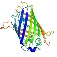
GFP has a typical beta barrel structure, consisting of one β-sheet with alpha helix(s) containing the chromophore running through the center.[4][5] Inward facing sidechains of the barrel induce specific cyclization reactions in the tripeptide Ser65–Tyr66–Gly67 that lead to chromophore formation. This process of post-translational modification is referred to as maturation. The hydrogen bonding network and electron stacking interactions with these sidechains influence the color of wtGFP and its numerous derivatives. The tightly packed nature of the barrel excludes solvent molecules, protecting the chromophore fluorescence from quenching by water.
[edit] History
[edit] Wild-type GFP (wtGFP)
In the 1960s and 1970s GFP, along with the separate luminescent protein aequorin, was first purified from Aequorea victoria and its properties studied by Osamu Shimomura.[6] In A. victoria, GFP fluorescence occurs when aequorin interacts with Ca2+ ions, inducing a blue glow. Some of this luminescent energy is transferred to the GFP, shifting the overall color towards green.[7] However, its utility as a tool for molecular biologists did not begin to be realized until 1992 when Douglas Prasher reported the cloning and nucleotide sequence of wtGFP in Gene.[8] The funding for this project had run out, so Prasher sent cDNA samples to several labs. The lab of Martin Chalfie expressed the coding sequence of wtGFP, with the first few amino acids deleted, in heterologous cells of E. coli and C. elegans, publishing the results in Science in 1994.[9] Frederick Tsuji's lab independently reported the expression of the recombinant protein one month later.[10] Remarkably, the GFP molecule folded and was fluorescent at room temperature, without the need for exogenous cofactors specific to the jellyfish. Although this near-wtGFP was fluorescent, it had several drawbacks, including dual peaked excitation spectra, pH sensitivity, chloride sensitivity, poor fluorescence quantum yield, poor photostability and poor folding at 37°C.
The first reported crystal structure of a GFP was that of the S65T mutant by the Remington group in Science in 1996.[4] One month later, the Phillips group independently reported the wild type GFP structure in Nature Biotech.[5] These crystal structures provided vital background on chromophore formation and neighboring residue interactions. Researchers have modified these residues by directed and random mutagenesis to produce the wide variety of GFP derivatives in use today. Martin Chalfie, Osamu Shimomura and Roger Y. Tsien share the 2008 Nobel Prize in Chemistry for their discovery and development of the green fluorescent protein.[11]
[edit] GFP derivatives

Due to the potential for widespread usage and the evolving needs of researchers, many different mutants of GFP have been engineered.[12] The first major improvement was a single point mutation (S65T) reported in 1995 in Nature by Roger Tsien.[13] This mutation dramatically improved the spectral characteristics of GFP, resulting in increased fluorescence, photostability and a shift of the major excitation peak to 488nm with the peak emission kept at 509 nm. This matched the spectral characteristics of commonly available FITC filter sets, increasing the practicality of use by the general researcher. A 37 °C folding efficiency (F64L) point mutant to this scaffold yielding enhanced GFP (EGFP) was discovered in 1995 by the lab of Ole Thastrup. [14] EGFP opened for using GFPs in mammalian cells. EGFP has an extinction coefficient (denoted ε) of 55,000 M−1cm−1.[15] The fluorescence quantum yield (QY) of EGFP is 0.60. The relative brightness, expressed as ε•QY, is 33,000 M−1cm−1. Superfolder GFP, a series of mutations that allow GFP to rapidly fold and mature even when fused to poorly folding peptides, was reported in 2006.[16]
Many other mutations have been made, including color mutants; in particular blue fluorescent protein (EBFP, EBFP2, Azurite, mKalama1), cyan fluorescent protein (ECFP, Cerulean, CyPet) and yellow fluorescent protein derivatives (YFP, Citrine, Venus, YPet). BFP derivatives (except mKalama1) contain the Y66H substitution. The critical mutation in cyan derivatives is the Y66W substitution, which causes the chromophore to form with an indole rather than phenol component. Several additional compensatory mutations in the surrounding barrel are required to restore brightness to this modified chromophore due to the increased bulk of the indole group. The red-shifted wavelength of the YFP derivatives is accomplished by the T203Y mutation and is due to π-electron stacking interactions between the substituted tyrosine residue and the chromophore.[2] These two classes of spectral variants are often employed for fluorescence resonance energy transfer (FRET) experiments. Genetically-encoded FRET reporters sensitive to cell signaling molecules, such as calcium or glutamate, protein phosphorylation state, protein complementation, receptor dimerization and other processes provide highly specific optical readouts of cell activity in real time.
Semirational mutagenesis of a number of residues led to pH-sensitive mutants known as pHluorins, and later super-ecliptic pHluorins. By exploiting the rapid change in pH upon synaptic vesicle fusion, pHluorins tagged to synaptobrevin have been used to visualize synaptic activity in neurons.[17]
Redox sensitive versions of GFP (roGFP) were engineered by introduction of cysteines into the beta barrel structure. The redox state of the cysteines determines the fluorescent properties of roGFP.[18]
The nomenclature of modified GFPs is often confusing due to overlapping mapping of several GFP versions onto a single name. For example, mGFP often refers to a GFP with an N-terminal palmitoylation that causes the GFP to bind to cell membranes. However, the same term is also used to refer to monomeric GFP, which is often achieved by the dimer interface breaking A206K mutation. Wild-type GFP has a weak dimerization tendency at concentrations above 5 mg/mL. mGFP also stands for "modified GFP" which has been optimized through amino acid exchange for stable expression in plant cells.
[edit] Use
The availability of GFP and its derivatives has thoroughly redefined fluorescence microscopy and the way it is used in cell biology and other biological disciplines.[19] While most small fluorescent molecules such as FITC (fluorescein isothiocyanate) are strongly phototoxic when used in live cells, fluorescent proteins such as GFP are usually much less harmful when illuminated in living cells. This has triggered the development of highly automated live cell fluorescence microscopy systems which can be used to observe cells over time expressing one or more proteins tagged with fluorescent proteins. For example, GFP had been widely used in labelling the spermatozoa of various organisms for identification purposes as in Drosophila melanogaster, where expression of GFP can be used as a marker for a particular characteristic. GFP can also be expressed in different structures enabling morphological distinction. In such cases, the gene for the production of GFP is spliced into the genome of the organism in the region of the DNA which codes for the target proteins, and which is controlled by the same regulatory sequence; that is the gene's regulatory sequence now controls the production of GFP, in addition to the tagged protein(s). In cells where the gene is expressed, and the tagged proteins are produced, GFP is produced at the same time. Thus, only those cells in which the tagged gene is expressed, or the target proteins are produced, will fluoresce when observed under fluorescence microscopy. Analysis of such time lapse movies has redefined the understanding of many biological processes including protein folding, protein transport, and RNA dynamics, which in the past had been studied using fixed (i.e. dead) material. .
Another powerful use of GFP is to express the protein in small sets of specific cells. This allows researchers to optically detect specific types of cells in vitro (in a dish), or even in vivo (in the living organism).[20] Genetically combining several spectral variants of GFP is a useful trick for the analysis of brain circuitry (Brainbow).
[edit] GFP in nature
The purpose of both bioluminescence and GFP fluorescence in jellyfish is unknown. GFP is co-expressed with aequorin in small granules around the rim of the jellyfish bell. The secondary excitation peak (480nm) of GFP does absorb some of the blue emission of aequorin, giving the bioluminescence a more green hue. The serine 65 residue of the GFP chromophore is responsible for the dual peaked excitation spectra of wild type GFP. It is conserved in all three GFP isoforms originally cloned by Prasher. Nearly all mutations of this residue consolidate the excitation spectra to a single peak at either 395nm or 480nm. The precise mechanism of this sensitivity is complex, but probably involves donation of a hydrogen from serine 65 to glutamate 222, which influences chromophore ionization.[2] Since a single mutation can dramatically enhance the 480nm excitation peak, making GFP a much more efficient partner of aequorin, A. victoria appears to evolutionarily prefer the less-efficient, dual peaked excitation spectrum. Roger Tsien has speculated that varying hydrostatic pressure with depth may effect serine 65's ability to donate a hydrogen to the chromophore and shift the ratio of the two excitation peaks. Thus the jellyfish may change the color of its bioluminescence with depth. Unfortunately, a collapse in the population of jellyfish in Friday Harbor, where GFP was originally discovered, has hampered further study of the role of GFP in the jellyfish's natural environment.
[edit] GFP in fine art

Julian Voss-Andreae, a German-born artist specializing in "protein sculptures,"[21] created sculptures based on the structure of GFP, including the 5'6" (1.70 m) tall "Green Fluorescent Protein" (2004)[22] and the 4'7" (1.40) tall "Steel Jellyfish" (2006). The latter sculpture is currently located at the place of GFP's discovery by Shimomura in 1962, the University of Washington's Friday Harbor Laboratories.[23]
[edit] Transgenic pets
Alba, a fluorescent rabbit, was commissioned by Eduardo Kac using GFP for purposes of art and social commentary.[24] The US company Yorktown Technologies markets green fluorescent zebrafish (GloFish) to aquarium shops. NeonPets, a US based company markets green fluorescent mice to the pet industry as NeonMice.[25]. Green fluorescent pigs, known as Noels were bred by a group of researchers led by Wu Shinn-Chih at the Department of Animal Science and Technology at National Taiwan University. [26]
[edit] References
- ^ Prendergast F, Mann K (1978). "Chemical and physical properties of aequorin and the green fluorescent protein isolated from Aequorea forskålea". Biochemistry 17 (17): 3448–53. doi:. PMID 28749.
- ^ a b c Tsien R (1998). "The green fluorescent protein" (PDF). Annu Rev Biochem 67: 509–44. doi:. PMID 9759496. http://tsienlab.ucsd.edu/Publications/Tsien%201998%20Annu.%20Rev.%20Biochem%20-%20GFP.pdf.
- ^ Phillips G (2001). "Green fluorescent protein--a bright idea for the study of bacterial protein localization". FEMS Microbiol Lett 204 (1): 9–18. doi:. PMID 11682170.
- ^ a b Ormö M, Cubitt A, Kallio K, Gross L, Tsien R, Remington S (1996). "Crystal structure of the Aequorea victoria green fluorescent protein". Science 273 (5280): 1392–5. doi:. PMID 8703075.
- ^ a b Yang F, Moss L, Phillips G (1996). "The molecular structure of green fluorescent protein". Nat Biotechnol 14 (10): 1246–51. doi:. PMID 9631087.
- ^ Shimomura O, Johnson F, Saiga Y (1962). "Extraction, purification and properties of aequorin, a bioluminescent protein from the luminous hydromedusan, Aequorea". J Cell Comp Physiol 59: 223–39. doi:. PMID 13911999.
- ^ Morise H, Shimomura O, Johnson F, Winant J (1974). "Intermolecular energy transfer in the bioluminescent system of Aequorea". Biochemistry 13 (12): 2656–62. doi:. PMID 4151620.
- ^ Prasher D, Eckenrode V, Ward W, Prendergast F, Cormier M (1992). "Primary structure of the Aequorea victoria green-fluorescent protein". Gene 111 (2): 229–33. doi:. PMID 1347277.
- ^ Chalfie M, Tu Y, Euskirchen G, Ward W, Prasher D (1994). "Green fluorescent protein as a marker for gene expression". Science 263 (5148): 802–5. doi:. PMID 8303295.
- ^ Inouye S, Tsuji F (1994). "Aequorea green fluorescent protein. Expression of the gene and fluorescence characteristics of the recombinant protein". FEBS Lett 341 (2-3): 277–80. doi:. PMID 8137953.
- ^ "The Nobel Prize in Chemistry 2008". 2008-10-08. http://nobelprize.org/nobel_prizes/chemistry/laureates/2008/. Retrieved on 2008-10-08.
- ^ Shaner N, Steinbach P, Tsien R (2005). "A guide to choosing fluorescent proteins" (PDF). Nat Methods 2 (12): 905–9. doi:. PMID 16299475. http://tsienlab.ucsd.edu/Publications/Shaner%202005%20Nature%20Methods%20-%20Choosing%20fluorescent%20proteins.pdf.
- ^ Heim R, Cubitt A, Tsien R (1995). "Improved green fluorescence" (PDF). Nature 373 (6516): 663–4. doi:. PMID 7854443. http://tsienlab.ucsd.edu/Publications/Heim%201995%20Nature%20-%20Improved%20GFP.PDF.
- ^ Thastrup O, Tullin S, Kongsbak Poulsen L, Bjørn S (1995). "Fluorescent Proteins". US patent. http://patft.uspto.gov/netacgi/nph-Parser?Sect2=PTO1&Sect2=HITOFF&p=1&u=%2Fnetahtml%2FPTO%2Fsearch-bool.html&r=1&f=G&l=50&d=PALL&RefSrch=yes&Query=PN%2F6172188.
- ^ Shelley R. McRae, Christopher L. Brown and Gillian R. Bushell (May 2005). "Rapid purification of EGFP, EYFP, and ECFP with high yield and purity". Protein Expression and Purification 41 (1): 121–127. doi:. PMID 15802229.
- ^ Pédelacq J, Cabantous S, Tran T, Terwilliger T, Waldo G (2006). "Engineering and characterization of a superfolder green fluorescent protein". Nat Biotechnol 24 (1): 79–88. doi:. PMID 16369541.
- ^ Miesenböck G, De Angelis D, Rothman J (1998). "Visualizing secretion and synaptic transmission with pH-sensitive green fluorescent proteins". Nature 394 (6689): 192–5. doi:. PMID 9671304.
- ^ Hanson GT, Aggeler R, Oglesbee D, Cannon M, Capaldi RA, Tsien RY, Remington SJ (2004). "Investigating mitochondrial redox potential with redox-sensitive green fluorescent protein indicators". J Biol Chem 279 (13): 13044-53. doi:. PMID 14722062.
- ^ Yuste R (2005). "Fluorescence microscopy today". Nat Methods 2 (12): 902–4. doi:. PMID 16299474.
- ^ Chudakov D, Lukyanov S, Lukyanov K (2005). "Fluorescent proteins as a toolkit for in vivo imaging". Trends Biotechnol 23 (12): 605–13. doi:. PMID 16269193.
- ^ Voss-Andreae, J (2005). "Protein Sculptures: Life's Building Blocks Inspire Art". Leonardo 38: 41–45. doi:.
- ^ Pawlak, Alexander (2005). "Inspirierende Proteine". Physik Journal 4: 12.
- ^ "Julian Voss-Andreae Sculpture". http://www.julianvossandreae.com/. Retrieved on 2007-06-14.
- ^ Eduardo Kac. "GFP Bunny". http://www.ekac.org/gfpbunny.html#gfpbunnyanchor.
- ^ [1] Glow-In-The Dark NeonMice
- ^ Scientists in Taiwan breed fluorescent green pigs
[edit] Further reading
- Pieribone V, Gruber D (2006). Aglow in the Dark: The Revolutionary Science of Biofluorescence. Cambridge: Belknap Press. ISBN 0674019210. OCLC 60321612. Popular science book describing history and discovery of GFP
- Zimmer M (2005). Glowing Genes: A Revolution In Biotechnology. Buffalo, NY: Prometheus Books. ISBN 1591022533. OCLC 56614624.
[edit] External links
- Video: Roger Tsien Gives Nobel Lecture at UC San Diego
- A comprehensive article on fluorescent protiens at Scholarpedia
- Introduction to fluorescent proteins
- History, uses, and structure of GFP
- Brief summary of landmark GFP papers
- Interactive Java applet demonstrating the chemistry behind the formation of the GFP chromophore
- Tsien Lab @ UCSD
- Video of 2008 Nobel Prize lecture of Roger Tsien on fluorescent proteins
- Excitation and emission spectra for various fluorescent proteins
- Timeline
- Some factoids about Aequorea, the jellyfish source of GFP
- Video introduction to GFP, from the "Secrets of the Sequence" educational video series
|
|||||||||||

