Vestibular system
From Wikipedia, the free encyclopedia
The vestibular system, which contributes to our balance and our sense of spatial orientation, is the sensory system that provides the dominant input about movement and equilibrioception. Together with the cochlea, a part of the auditory system, it constitutes the labyrinth of the inner ear, situated in the vestibulum in the inner ear (Figure 1). As our movements consist of rotations and translations, the vestibular system comprises two components: the semicircular canal system, which indicate rotational movements; and the otoliths, which indicate linear accelerations. The vestibular system sends signals primarily to the neural structures that control our eye movements, and to the muscles that keep us upright. The projections to the former provide the anatomical basis of the vestibulo-ocular reflex, which is required for clear vision; and the projections to the muscles that control our posture are necessary to keep us upright.

Contents |
[edit] Semicircular canal system
The semicircular canal system detects rotational movements. The semicircular canals are its main tools to achieve this detection.
[edit] Structure
As the basis of our perception of a three-dimensional world, our vestibular system contains three semicircular canals in each labyrinth. They are approximately orthogonal to each other, and are called the horizontal (or lateral), the anterior semicircular canal (or superior) and the posterior (or inferior) semicircular canal. Anterior and posterior canals may be collectively called vertical semicircular canals.
- Movement of fluid within the horizontal semicircular canal corresponds to rotation of the head around a vertical axis (i.e. the neck), as when doing a pirouette.
- The anterior and posterior semicircular canals detect rotations of the head in the sagittal plane (as when nodding), and in the frontal plane, as when cartwheeling. Both anterior and posterior canals are oriented at approximately 45° between frontal and sagittal planes.
The movement of fluid pushes on a structure called cupula, which contains hair cells that transduct the mechanical movement to electrical signals [1]
[edit] Push-pull systems

The canals are arranged in such a way that each canal on the left side has an almost parallel counterpart on the right side. Each of these three pairs works in a push-pull fashion: when one canal is stimulated, its corresponding partner on the other side is inhibited, and vice versa.
This push-pull system allows us to sense all directions of rotation: while the right horizontal canal gets stimulated during head rotations to the right (Fig 2), the left horizontal canal gets stimulated (and thus predominantly signals) by head rotations to the left.
Vertical canals are coupled in a crossed fashion, i.e. stimulations that are excitatory for an anterior canal are also inhibitory for the contralateral posterior, and vice versa.
[edit] Vestibulo-ocular reflex (VOR)
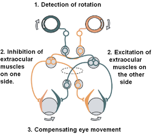
The vestibulo-ocular reflex (VOR) is a reflex eye movement that stabilizes images on the retina during head movement by producing an eye movement in the direction opposite to head movement, thus preserving the image on the center of the visual field. For example, when the head moves to the right, the eyes move to the left, and vice versa. Since slight head movements are present all the time, the VOR is very important for stabilizing vision: patients whose VOR is impaired find it difficult to read, because they cannot stabilize the eyes during small head tremors. The VOR reflex does not depend on visual input and works even in total darkness or when the eyes are closed.
This reflex, combined with the push-pull principle described above, forms the physiological basis of the Rapid head impulse test or Halmagyi-Curthoys-test, in which the head is rapidly and forcefully moved to the side, while controlling if the eyes keep looking in the same direction.
[edit] Mechanics
The mechanics of the semicircular canals can be described by a damped oscillator. If we designate the deflection of the cupula with θ, and the head velocity with  , the cupula deflection is approximately

α is a proportionality factor, and s corresponds to the frequency. For humans, the time constants T1 and T2 are approximately 3 ms and 5 s, respectively. As a result, for typical head movements, which cover the frequency range of 0.1 Hz and 10 Hz, the deflection of the cupula is approximately proportional to the head-velocity. This is very useful, since the velocity of the eyes must be opposite to the velocity of the head in order to have clear vision.
[edit] Central processing
Signals from the vestibular system also project to the cerebellum (where they are used to keep the VOR effective, a task usually referred to as learning or adaptation) and to different areas in the cortex. The projections to the cortex are spread out over different areas, and their implications are currently not clearly understood.
[edit] Otolithic organs
While the semicircular canals respond to rotations, the otolithic organs sense linear accelerations. We have two on each side, one called utricle, the other saccule. The otoconia crystals in the otoconia layer rest on a viscous gel layer, and are heavier than their surroundings. Therefore they get displaced during linear acceleration, which in turn deflects the ciliary bundles of the hair cells and thus produces a sensory signal. Most of the utricular signals elicit eye movements, while the majority of the saccular signals projects to muscles that control our posture. While the interpretation of the rotation signals from the semicircular canals is straightforward, the interpretation of otolith signals is more difficult: since gravity is equivalent to a constant linear acceleration, we somehow have to distinguish otolith signals that are caused by linear movements from such that are caused by gravity. We can do that quite well, but the neural mechanisms underlying this separation are not yet fully understood.
[edit] Experience from the vestibular system

Experience from the vestibular system is called equilibrioception. It is mainly used for the sense of balance and for spatial orientation. When the vestibular system is stimulated without any other inputs, one experiences a sense of self motion. For example, a person in complete darkness and sitting in a chair will feel that he or she has turned to the left if the chair is turned to the left. A person in an elevator, with essentially constant visual input, will feel she is descending as the elevator starts to descend.
[edit] Vestibular/somatogyral illusions
See Sensory illusions in aviation.
[edit] Pathologies
Diseases of the vestibular system can take different forms, and usually induce vertigo and instability, often accompanied by nausea. The most common ones are Vestibular neuritis, a related condition called Labyrinthitis, and BPPV. In addition, the function of the vestibular system can be affected by tumors on the cochleo-vestibular nerve, an infarct in the brain stem or in cortical regions related to the processing of vestibular signals, and cerebellar atrophy.
Alcohol can also cause alterations in the vestibular system for short periods of time and will result in vertigo and possibly nystagmus. This is due to the variable viscosity of the blood and the endolymph during the consumption of alcohol. The common term for this type of sensation is the "Bed Spins".
- PAN I - The alcohol concentration is higher in the blood than in the vestibular system, hence the endolymph is relatively dense.
- PAN II - The alcohol concentration is lower in the blood than in the vestibular system, hence the endolymph is relatively dilute.
It is interesting to note that PAN I will result in subjective vertigo in one direction and typically occurs shortly after ingestion of alcohol when blood alcohol levels are highest. PAN II will eventually cause subjective vertigo in the opposite direction. This occurs several hours after ingestion and after a relative reduction in blood alcohol levels.
[edit] BPPV
BPPV, which is short for Benign Paroxysmal Positional Vertigo, is probably caused by pieces that have broken off from the Otoliths, and have slipped into one of the semicircular canals. In most cases it is the posterior canal that is affected. In certain head positions, these particles shift and create a fluid wave which displaces the cupula of the canal affected, which leads to dizziness, vertigo and nystagmus.
[edit] External links
- SensesWeb, which has been created by Tutis Vilis, contains animations – of all sensory systems, as well as the corresponding PDF files, and additional further links.
- Dizzytimes.com Online Community for Sufferers of Vertigo and Dizziness.
[edit] Footnotes
- ^ Medical Physiology, Walter Boron & Emile Boulpaep, ISBN 1-4160-2328-3, Elsevier Saunders 2005. Updated edition. 1300 pages.
[edit] References
- S. M. Highstein, R. R. Fay, A. N. Popper, editors (2004). The vestibular system. Berlin: Springer. ISBN 0-387-98314-7. (Comment: A book for experts, summarizing the state of the art in our understanding of the balance system)
- Thomas Brandt (2003). Vertigo : Its Multisensory Syndromes. Berlin: Springer. ISBN 0-387-40500-3. (Comment: For clinicians, and other professionals working with dizzy patients.)
- Driver Drowsiness: Is something missing? J. Christopher Brill, Peter A. Hancock, Richard D. Gilson. University of Central Florida (2003) link (Comment: Research on driver or motion-induced sleepiness aka 'sopite syndrome' links it to the vestibular labyrinths.)
|
|||||||||||
|
||||||||||||||||||||

