Prenatal development
From Wikipedia, the free encyclopedia
- "Unborn child" redirects here; for other uses, see unborn child (disambiguation).
Prenatal or antenatal development is the process in which an embryo or fetus (or foetus) gestates during pregnancy, from fertilization until birth. Often, the terms fetal development, foetal development, or embryology are used in a similar sense.
After fertilization the embryogenesis starts. In humans, when embryogenesis finishes, by the end of the 10th week of gestational age, the precursors of all the major organs of the body have been created. Therefore, the following period, the fetal period, is described both topically on one hand, i.e. by organ, and strictly chronologically on the other, by a list of major occurrences by weeks of gestational age.
[edit] Fertilization

When semen is deposited in the vagina, the spermatozoa travel through the cervix and body of the uterus and into the Fallopian tubes. Fertilization of the ovum (egg cell) usually takes place in the Fallopian tube. Many sperm must cooperate to penetrate the thick protective shell-like barrier that surrounds the ovum. The first sperm that penetrates fully into the egg donates its genetic material (DNA). The egg then polarizes, repelling any additional sperm. The resulting combination is called a zygote. The term "conception" refers variably to either fertilization or to formation of the conceptus after uterine implantation, and this terminology is controversial.
Prior to fertilization, each ovum contains a complete human genome, including a single X but no Y chromosome. Likewise, each spermatozoon contains a complete set of autosomes and a single sex chromosome, either X or Y. The resulting zygote is similar to the majority of somatic cells because it contains two copies of the genome in a diploid set of chromosomes. One set of chromosomes came from the nucleus of the ovum and the second set from the nucleus of the sperm. If the spermatozoon contributes a Y chromosome then the zygote will develop as a male. Unlike the X chromosome, the Y chromosome contains very little genetic information. However it does contain a gene, SRY, which will switch on androgen production at a later stage, leading to the development of a male body type. In contrast, the mitochondrial genetic information of the zygote comes entirely from the mother via the ovum.
[edit] Embryonic period
The embryonic period in humans begins at fertilization (2nd week of gestation) and continues until the end of the 10th week of gestation (8th week of development).
The zygote spends the next few days traveling down the Fallopian tube. Meanwhile it divides several times to form a ball of cells called a morula. Further cellular division is accompanied by the formation of a small cavity between the cells. This stage is called a blastocyst. Up to this point there is no growth in the overall size of the embryo, so each division produces successively smaller cells.
The blastocyst reaches the uterus at roughly the fifth day after fertilization. It is here that lysis of the zona pellucida, a glycoprotein shell, occurs. This is required so that the trophectoderm cells, which give rise to extra-embryonic structures such as the placenta, of the blastocyst can come into contact with the luminal epithelial cells of the endometrium. (Contrast this with zona hatching, an event that occurs in vitro by a different mechanism, but with a similar result). It then adheres to the uterine lining and becomes embedded in the endometrial cell layer. This process is also called implantation. In most successful pregnancies, the conceptus implants 8 to 10 days after ovulation (Wilcox et al 1999). The inner cell mass forms the embryo, while the outer cell layers form the membranes and placenta. Together, the embryo and its membranes are referred to as a conceptus, or the "products of conception".
Rapid growth occurs and the embryo's main external features begin to take form. This process is called differentiation, which produces the varied cell types (such as blood cells, kidney cells, and nerve cells). A spontaneous abortion, or miscarriage, in the first trimester of pregnancy is usually due to major genetic mistakes or abnormalities in the developing embryo. During this critical period (most of the first trimester), the developing embryo is also susceptible to toxic exposures, such as:
- Alcohol, certain drugs, and other toxins that cause birth defects, such as Fetal alcohol syndrome
- Infection (such as rubella or cytomegalovirus)
- Radiation from x-rays or radiation therapy
- Nutritional deficiencies such as lack of folate which contributes to spina bifida
Generally, if a structure pre-dates another structure in evolutionary terms, then it often appears earlier than the other in an embryo; this general observation is sometimes summarized by the phrase "ontogeny recapitulates phylogeny."[1] For example, the backbone is a common structure among all vertebrates such as fish, reptiles and mammals, and the backbone also appears as one of the earliest structures laid out in all vertebrate embryos. The cerebrum in humans, which is the most sophisticated part of the brain, develops last. The concept of recapitulation is not absolute, but it is recognized as being partly applicable to development of the human embryo.[1]
[edit] Changes by weeks of gestational age
[edit] Week 1 (day 1-7 of fertilization)
- Fertilization of the ovum to form a zygote. (day 1 of fert.[2])
- The zygote undergoes mitotic cellular divisions, but does not increase in size. This mitosis is also known as cleavage. A hollow cavity forms marking the blastocyst stage. (day 1.5-3 of fert.[2])
- The blastocyst contains only a thin rim of trophoblast cells and a clump of cells at one end known as the "embryonic pole" which include embryonic stem cells.
- The embryo hatches from its protein shell (zona pellucida) and performs implantation onto the endometrial lining of the mother's uterus. (day 5-6 of fert.[2])
- If separation into identical twins occurs, 1/3 of the time it will happen before day 5.[3]
[edit] Week 2 (8-14 days from fertilization)
- Trophoblast cells surrounding the embryonic cells proliferate and invade deeper into the uterine lining. They will eventually form the placenta and embryonic membranes. The blastocyst is fully implanted day 7-12 of fert. [2]
- Formation of the yolk sac.
- The embryonic cells flatten into a disk, two cells thick.
- If separation into identical twins occurs, 2/3 of the time it will happen between days 5 and 9. If it happens after day 9, there is a significant risk of the twins being conjoined.
- Primitive streak develops. (day 13 of fert.[2])
- Primary stem villi appear. (day 13 of fert.[2])
[edit] Week 3 (15-21 days from fertilization)
- A notochord forms in the center of the embryonic disk. (day 16 of fert.[2])
- Gastrulation commences. (day 16of fert.[2])
- A neural groove (future spinal cord) forms over the notochord with a brain bulge at one end. Neuromeres appear. (day 18 of fert.[2])
- Somites, the divisions of the future vertebra, form. (day 20 of fert.[2])
- Primitive heart tube is forming. Vasculature begins to develop in embryonic disc. (day 20 of fert.[2])

[edit] Week 4 (22-28 days from fertilization)
- The embryo measures 4 mm (1/8 inch) in length and begins to curve into a C shape.
- The heart bulges, further develops, and begins to beat in a regular rhythm. Septum primum appear. [2]
- Branchial arches, grooves which will form structures of the face and neck, form.
- The neural tube closes.
- The ears begin to form as otic pits.
- Arm buds and a tail are visible.
- Pulmonary primordium, the first traits of the lung appear. [2]
- Hepatic plate, the first traits of the liver appear. [2]
- Buccopharyngeal membrane ruptures. This is the future mouth. [2]
- Cystic diverticulum, which will become the gallbladder, and dorsal pancreatic bud, which will become the pancreas appear. [2]
- Urorectal septum begins to form. Thus, the rectal and urinary passageways become separated. [2]
- Anterior and posterior horns differentiate in the spinal cord [2]
- Spleen appears. [2]
- Ureteric buds appear. [2]
[edit] Week 5 (29-35 days from fertilization)
- The embryo measures 8 mm (1/4 inch) in length.
- Lens pits and optic cups form the start of the developing eye.
- Nasal pits form.
- The brain divides into 5 vesicles, including the early telencephalon.
- Leg buds form and hands form as flat paddles on the arms.
- Rudimentary blood moves through primitive vessels connecting to the yolk sac and chorionic membranes.
[edit] Week 6 (36-42 days from fertilization)
- The embryo measures 13 mm (1/2 inch) in length.
- Lungs begin to form.
- The brain continues to develop.
- Arms and legs have lengthened with foot and hand areas distinguishable.
- The hands and feet have digits, but may still be webbed.
- The gonadal ridge begins to be perceptible.
- The lymphatic system begins to develop.
[edit] Week 7 (43-49 days from fertilization)
- The embryo measures 18 mm (3/4 inch) in length.
- Nipples and hair follicles begin to form.
- Location of the elbows and toes are visible.
- Spontaneous limb movements may be detected by ultrasound.
- All essential organs have at least begun formation.
[edit] Fetal period
The fetal period begins at the end of the 10th week of gestation (8th week of development). Since the precursors of all the major organs are created by this time, the fetal period is described both by organ and by a list of changes by weeks of gestational age.
Because the precursors of the organs are formed, fetus also is not as sensitive to damage from environmental exposures as the embryo. Instead, toxic exposures often cause physiological abnormalities or minor congenital malformation.
[edit] By organ
Each organ has its own development.
- Development of circulatory system
- Development of digestive system
- Development of endocrine system
- Development of integumentary system
- Development of lymphatic system
- Development of muscular system
- Development of nervous system
- Development of the urinary and reproductive system
- Development of respiratory system
[edit] Changes by weeks of gestational age
See also: Human fetal development
From the 8th week until birth (around 38 weeks), the developing organism is called a fetus. The fetus is not as sensitive to damage from environmental exposures as the embryo, and toxic exposures often cause physiological abnormalities or minor congenital malformation. All major structures are already formed in the fetus, but they continue to grow and develop.
[edit] Week 8 (7th week of development)
- Embryo measures 30 mm (1.2 inches) in length.
- Intestines rotate.
- Facial features continue to develop.
- the eyelids are more developed.
- the external features of the ear begin to take their final shape.
[edit] Weeks 9 to 12 (8th to 11th week of development)
- The fetus reaches a length of 8 cm (3.2 inches).
- The head comprises nearly half of the fetus' size.
- The face is well formed
- The eyelids close and will not reopen until about the 28th week.
- Tooth buds, which will form the baby teeth, appear.
- The limbs are long and thin.
- The fetus can make a fist with its fingers.
- Genitals appear well differentiated.
- Red blood cells are produced in the liver.
[edit] Weeks 13 to 16 (12th to 15th week of development)
- The fetus reaches a length of about 15 cm (6 inches).
- A fine hair called lanugo develops on the head.
- Fetal skin is almost transparent.
- More muscle tissue and bones have developed, and the bones become harder.
- The fetus makes active movements.
- Sucking motions are made with the mouth.
- Meconium is made in the intestinal tract.
- The liver and pancreas produce fluid secretions.

[edit] Week 19 (18th week of development)
- The fetus reaches a length of 20 cm (8 inches).
- Lanugo covers the entire body.
- Eyebrows and eyelashes appear.
- Nails appear on fingers and toes.
- The fetus is more active with increased muscle development.
- "Quickening" usually occurs (the mother can feel the fetus moving).
- The fetal heartbeat can be heard with a stethoscope.
[edit] Week 23 (22nd week of development)
- The fetus reaches a length of 28 cm (11.2 inches).
- The fetus weighs about 725 g (1 lb 10 oz).
- Eyebrows and eyelashes are well formed.
- All of the eye components are developed.
- The fetus has a hand and startle reflex.
- Footprints and fingerprints continue forming.
- Alveoli (air sacs) are forming in lungs.
[edit] Week 27 (26th week of development)
- The fetus reaches a length of 38 cm (15 inches).
- The fetus weighs about 1.2 kg (2 lb 11 oz).
- The brain develops rapidly.
- The nervous system develops enough to control some body functions.
- The eyelids open and close.
- The cochleae are now developed, though the myelin sheaths in neural portion of the auditory system will continue to develop until 18 months after birth.
- The respiratory system, while immature, has developed to the point where gas exchange is possible.
[edit] Week 31 (30th week of development)
- The fetus reaches a length of about 38-43 cm (15-17 inches).
- The fetus weighs about 2 kg (3 lb 0 oz).
- The amount of body fat rapidly increases.
- Rhythmic breathing movements occur, but lungs are not fully mature.
- Thalamic brain connections, which mediate sensory input, form.
- Bones are fully developed, but are still soft and pliable.
- The fetus begins storing iron, calcium, and phosphorus.
[edit] Week 35 (34th week of development)
- The fetus reaches a length of about 40-48 cm (16-19 inches).
- The fetus weighs about 2.5 to 3 kg (5 lb 12 oz to 6 lb 12 oz).
- Lanugo begins to disappear.
- Body fat increases.
- Fingernails reach the end of the fingertips.
- a baby born at 36 weeks has a high chance of survival, but may require medical interventions.
[edit] Weeks 36 to 39 (35th to 38th week of development)
- The fetus is considered full-term at the end of the 37th week of gestational age.
- It may be 48 to 53 cm (19 to 21 inches) in length.
- The lanugo is gone except on the upper arms and shoulders.
- Fingernails extend beyond fingertips.
- Small breast buds are present on both sexes.
- Head hair is now coarse and thickest.
The development is continued postnatally with child development stages.
[edit] References
- ^ a b Stephen Jay Gould,. Ontogeny and Phylogeny. Cambridge, Mass: Belknap Press. p. 206. ISBN 0-674-63941-3. http://www.sjgarchive.org/library/ontogeny.html.
- ^ a b c d e f g h i j k l m n o p q r s t )William J. Larsen (2001). Human embryology. Edinburgh: Churchill Livingstone. ISBN 0-443-06583-7.
- ^ Scott F. Gilbert; with a chapter on plant development by Susan R. Singer (2000). Developmental biology. Sunderland, Mass: Sinauer Associates. ISBN 0-87893-243-7. http://www.ncbi.nlm.nih.gov/books/bv.fcgi?rid=dbio.box.2669.
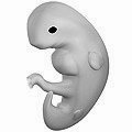
- ^ 3D Pregnancy (large image of fetus at 4 weeks after fertilization). Retrieved 2007-08-28. A rotatable 3D version of this photo is available here, and a sketch is available here.
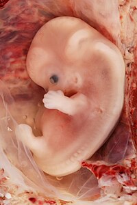
- ^ 3D Pregnancy (large image of fetus at 10 weeks after fertilization). Retrieved 2007-08-28. A rotatable 3D version of this photo is available here, and a sketch is available here.
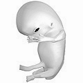
- ^ 3D Pregnancy (large image of fetus at 18 weeks after fertilization). Retrieved 2007-08-28. A rotatable 3D version of this photo is available here, and a sketch is available here.
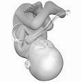
- ^ 3D Pregnancy (large image of fetus at 38 weeks after fertilization). Retrieved 2007-08-28. A rotatable 3D version of this photo is available here, and a sketch is available here.
- "MedlinePlus Medical Encyclopedia"
- Moore, Keith L. The Developing Human: 3rd Edition. W.B. Saunders Company, Philadelphia PA
- Wilcox AJ, Baird DD, Weinberg CR. Time of implantation of the conceptus and loss of pregnancy. 1999 N Engl J Med. 340(23):1796-9. PMID 10362823
- Ljunger, E, Cnattingius, S, Lundin, C, & Annerén, G. 2005 Chromosomal anomalies in first-trimester miscarriages. Acta Obstetricia et Gynecologica Scandinavica 84(11):1103-1107. PMID 10362823
[edit] See also
[edit] External links
| Wikimedia Commons has media related to: Embryology |
- Fetal Development Timeline from AboutKidsHealth.ca
- The Changes in Each Stage of Human Development
- Real Time Presentation of Human Embryo Development
|
||||||||||||||||||||||||||||||||||||||||||||||||||||||||||
|
|||||||||||||||||||||||
|
|||||||||||||||||||||||||||||||||||||||||||||
|
||||||||||||||||||||||||||||||||||||

