Sarcoidosis
From Wikipedia, the free encyclopedia
| Sarcoidosis Classification and external resources |
|
 |
|
|---|---|
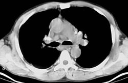
| Sarcoidosis in a Lymph Node. | |
| ICD-10 | D86. |
| ICD-9 | 135 |
| OMIM | 181000 |
| DiseasesDB | 11797 |
| MedlinePlus | 000076 |
| eMedicine | med/2063 |
| MeSH | D012507 |
Sarcoidosis (sarcoid = sarcoma-like, -osis = a process), also called sarcoid or Besnier-Boeck disease, is a multisystem disorder characterized by non-caseating granulomas (small inflammatory nodules). The cause of the disease is still unknown. Virtually any organ can be affected; however, granulomas most often appear in the lungs or the lymph nodes. Symptoms usually appear gradually but can occasionally appear suddenly. The clinical course generally varies and ranges from asymptomatic disease to a debilitating chronic condition that may lead to death.
Contents |
[edit] Epidemiology
Sarcoidosis most commonly affects young adults of both sexes, with a slight preponderance for women having been reported by most studies. Incidence is highest for individuals younger than 40 and peaks in the age-group from 20 to 29 years, a second peak is observed for women over 50[1] [2] .
Sarcoidosis occurs throughout the world in all races with an average incidence of 16.5/100,000 in men and 19/100,000 in women. The disease is most prevalent in Northern European countries, and the highest annual incidence of 60 per 100,000 is found in Sweden and Iceland. In the United States, sarcoidosis is more common in people of African descent than Caucasians, with annual incidence reported as 35.5 and 10.9 per 100,000, respectively.[3] Sarcoidosis is less commonly reported in South America, Spain and India, Canada and Philippines. In particular, Sarcoidosis is almost never reported in immigrants from northern Spain.
The differing incidence across the world may be at least partially attributable to the lack of screening programs in certain regions of the world and the overshadowing presence of other granulomatous diseases such as tuberculosis, that may interfere with the diagnosis of sarcoidosis where they are prevalent.[1]
There may also be racial differences in the severity of the disease. Several studies suggest that the presentation in people of African origin may be more severe and disseminated than for Caucasians, who are more likely to have asymptomatic disease.[4]
Manifestation appears to be slightly different according to race and gender, erythema nodosum is far more common in men than women and Caucasians than other races. In Japanese ophthalmologic and cardiac involvement is more common than in other races[2].
Sarcoidosis is one of the few pulmonary diseases with a higher prevalence in non-smokers.
[edit] Signs and symptoms
Sarcoidosis is a systemic disease that can affect any organ. Common symptoms are vague, such as fatigue unchanged by sleep, lack of energy, weight loss, aches and pains, arthralgia, dry eyes, blurry vision, shortness of breath, a dry hacking cough or skin lesions. The cutaneous symptoms vary, and range from rashes and noduli (small bumps) to erythema nodosum or lupus pernio. It is often asymptomatic.
The combination of erythema nodosum, bilateral hilar lymphadenopathy and arthralgia is called Löfgren syndrome. This syndrome has a relatively good prognosis.
Renal, liver (including portal hypertension), heart[5] or brain involvement may cause further symptoms and altered functioning. Manifestations in the eye include uveitis, uveoparotitis, and retinal inflammation, which may result in loss of visual acuity or blindness. Sarcoidosis affecting the brain or nerves is known as neurosarcoidosis.
The combination of anterior uveitis, parotitis, VII cranial nerve paralysis and fever is called uveoparotitid fever, and is associated with Heerfordt-Waldenstrom syndrome. (D86.8)
Sarcoidosis of the scalp presents with diffuse or patchy hair loss.[6]
[edit] Investigations
Hypercalcemia (high calcium levels) and its symptoms may be the result of excessive conversion of vitamin D to its active form by epithelioid macrophages.
Sarcoidosis most often manifests as a restrictive disease of the lungs, causing a decrease in lung volume and decreased compliance (the ability to stretch). The disease typically limits the amount of air drawn into the lungs, but produces higher than normal expiratory flow ratios. The vital capacity (full breath in, to full breath out) is decreased, and most of this air can be blown out in the first second. This means the FEV1/FVC ratio is increased from the normal of about 80%, to 90%. Obstructive lung changes, causing a decrease in the amount of air that can be exhaled, may occur when enlarged lymph nodes in the chest compress airways or when internal inflammation or nodules impede airflow.
Chest X-ray changes are divided into four stages
- Stage 1 bihilar lymphadenopathy
- Stage 2 bihilar lymphadenopathy and reticulonodular infiltrates
- Stage 3 bilateral pulmonary infiltrates
- Stage 4 fibrocystic sarcoidosis typically with upward hilar retraction, cystic & bullous changes
Because sarcoidosis can affect multiple organ systems, follow-up on a patient with sarcoidosis should always include an electrocardiogram, ocular examination by an ophthalmologist, liver function tests, serum calcium and 24-hour urine calcium. In female patients sarcoidosis is significantly associated with hypothyroidism, hyperthyroidism and other thyroid diseases, hence close surveillance of thyroid function is recommended [7] .
[edit] Causes and pathophysiology
The exact cause of sarcoidosis is not known. The current working hypothesis is that in genetically susceptible individuals sarcoidosis is caused through alteration in immune response after exposure to an environmental, occupational, or infectious agent.[8]
[edit] Anergy
On the one hand the disease is characterised by increased macrophage and CD4 helper T-cell activation resulting in accelerated inflammation – on the other hand the immune system of patients shows suppressed response to antigen challenges such as tuberculin. This paradoxic state of hyper- and hypo- activity at the same time is suggestive of a state of anergy. The anergy may also be responsible for the increased risk of infections and cancer. It appears that regulatory T-lymphocytes in the periphery of sarcoid granulomas suppress IL-2 secretion which is hypothesized to cause the state of anergy by preventing antigen-specific memory responses.[9]
While it is widely believed that TNF-alpha plays an important role in the formation of granulomas it was observed that sarcoidosis can be triggered by treatment with the TNF-alpha antagonist etanercept.[10][11]
[edit] Genetic associations
Investigations of genetic susceptibility yielded many candidate genes but only few were confirmed by further investigations and no reliable genetic markers are known. Currently most interesting candidate gene is BTNL2, several HLA-DR risk alleles are also investigated.[12] In persistent sarcoidosis the HLA haplotype HLA-B7-DR15 are either cooperating in disease or another gene between these two loci is associated. In non-persistent disease there is a strong genetic association with HLA DR3-DQ2.[13] Siblings have only a modestly increased risk (hazard ratio 5-6) to develop the disease, indicating that genetic susceptibility plays only a small role. The alternate hypothesis that family members share similar exposures to environmental pathogens is quite plausible to explain the apparent hereditary factor.
[edit] Infectious agents
Several infectious agents appear to be significantly associated with sarcoidosis but none of the known associations is specific enough to suggest a direct causative role. Propionibacterium acnes can be found in bronchoalveolar lavage of approximately 70% patients and is associated with disease activity, however it can be also found in 23% of controls.[14][15] A recent meta-analysis investigating the role of mycobacteria in sarcoidosis found it was present in 26.4% of cases, however the meta-analysis also detected a possible publication bias, so the results need further confirmation.[16][17]
There have also been reports of transmission of sarcoidosis via organ transplants.[18]
[edit] Vitamin D dysregulation
Sarcoidosis frequently causes a dysregulation of vitamin D production with an increase in extrarenal (outside the kidney) production.[19] Specifically, macrophages inside the granulomas convert vitamin D to its active form, resulting in elevated levels of the hormone 1,25-dihydroxyvitamin D and symptoms of hypervitaminosis D that may include fatigue, lack of strength or energy, irritability, metallic taste, temporary memory loss or cognitive problems. Physiological compensatory responses (e.g. suppression of the parathyroid hormone levels) may mean the patient does not develop frank hypercalcemia. This condition may be aggravated by high levels of estradiol and prolactin such as in pregnancy, leading to hypercalciuria and/or compensatory hypoparathyroidism.[20] High levels of Vitamin D are also implicated in immune-system dysfunctions which tie into the sarcoid condition.
The connection between Vitamin D disregulation, its effects on immune function, and how the combined effect might mediate sarcoidosis, is currently being explored by Dr. Trevor Marshall. The theory which Dr. Marshall is currently researching via a protocol (the Marshall Protocol) under FDA oversight, asserts that L-form or cell wall deficient bacteria infect the body, and evade the immune system by inducing or enhancing this Vitamin D disregulation, which in turn suppresses the body's immune response to said bacterial infection.
[edit] Hyperprolactinemia
Prolactin is frequently increased in sarcoidosis, between 3–32% cases have hyperprolactinemia [21], this frequently leads to amenorrhea, galactorrhea or nonpuerperal mastitis in women. Prolactin also has a broad spectrum of effects on the immune system and increased prolactin levels are associated with disease activity or may exacerbate symptoms in many autoimmune diseases and treatment with prolactin lowering medication has been shown effective in some cases. [22] However it is unknown if this relation holds in sarcoidosis and the gender predilection in sarcoidosis is less pronounced than in some other autoimmune diseases where such relation has been established. In pregnancy the effects of prolactin and estrogen counteract each other to some degree, with a slight trend to improve pulmonary manifestations of sarcoidosis while lupus, uveitis and arthralgia might slightly worsen[20]. Lupus, uveitis and arthralgia are known to be in some cases associated with increased prolactin levels and respond to bromocriptin treatment but so far this has not been investigated specifically for sarcoidosis. The reasons for increased prolactin levels in sarcoidosis are hitherto uncertain. It has been observed that prolactin is produced by T-lymphocytes in some autoimmune disorders in amounts high enough to affect the feedback by the hypothalamic dopaminergic system [23] . The extrapituitary prolactin is believed to play a role as a cytokine like proinflammatory factor. Prolactin antibodies are believed to play a role in hyperprolactinemia in other autoimmune disorders and high prevalence endocrine autoimmunity has been observed in patients with sarcoidosis [24] . It may also be a consequence of renal disease or treatment with steroids. Neurosarcoidosis may occasionally cause hypopituiarism but has not been reported to cause hyperprolactinemia.
[edit] Thyroid disease
In women a substantial association of thyroid disease and sarcoidosis has been reported. The association is less marked but still significant for male patients. Female patients have a significantly elevated risk for hypothyroidism, hyperthyroidism and thyroid autoimmunity and it appears that autoimmunity is very important in the pathogenesis of thyroid disease in this population. Thyroid granulomatosis on the other hand is uncommon [7] .
[edit] Hypersensitivity/autoimmune
Association of autoimmune disorders has been frequently observed. The exact mechanism of this relation is not known but some evidence supports the hypothesis that this is a consequence of Th1 lymphokine prevalence[25] [7].
Sarcoidosis has been associated with celiac disease. Celiac disease is a condition in which there is a chronic reaction to certain protein chains, commonly referred to as glutens, found in some cereal grains. This reaction causes destruction of the villi in the small intestine, with resulting malabsorption of nutrients.
An association with type IV hypersensitivity has been described.[26] Tests of delayed cutaneous hypersensitivity have been used to measure progression.[27]
[edit] Other
While disputed, some cases have been associated with inhalation of the dust from the collapse of the World Trade Center after the September 11, 2001 attacks.[28] See Health effects arising from the September 11, 2001 attacks for more information.
[edit] Pregnancy
Sarcoidosis generally does not prevent successful pregnancy and delivery, the endogenous estrogen in pregnancy may even have a slightly beneficial immunomodulatory effect. In most cases the course of sarcoidosis is unaffected by pregnancy, there is improvement in a few cases and worsening of symptoms in very few cases[20].
[edit] Treatment
Between 30 to 70% of patients do not require therapy[2].Corticosteroids, most commonly prednisone, have been the standard treatment for many years. In some patients, this treatment can slow or reverse the course of the disease, but other patients do not respond to steroid therapy. The use of corticosteroids in mild disease is controversial because in many cases the disease remits spontaneously. [29] Additionally, corticosteroids have many recognized dose- and duration-related side effects (which can be reduced through the use of alternate-day dosing for those on chronic prednisone therapy [30]), and their use is generally limited to severe, progressive, or organ-threatening disease. The influence of corticosteroids or other immunosuppressants on the natural history is unclear.
Severe symptoms are generally treated with steroids, and steroid-sparing agents such as azathioprine and methotrexate are often used. Rarely, cyclophosphamide has also been used. As the granulomas are caused by collections of immune system cells, particularly T cells, there has been some early indications of success using immunosuppressants, interleukin-2 inhibitors or anti-tumor necrosis factor-alpha treatment (such as infliximab). Unfortunately, none of these has provided reliable treatment, and there can be significant side effects such as an increased risk of reactivating latent tuberculosis. Anti-tumor necrosis factor-alpha treatment with etanercept in rheumatoid arthritis has been observed to actually cause sarcoidosis [10] .
The connection between vitamin D dysregulation and the emergence of sarcoidosis is being explored under the Marshall Protocol, which asserts that cell-wall deficient bacteria infect the body and evade immune surveillance by promoting such dysregulation, which in turn suppresses the body's immune response.
[edit] Prognosis
Approximately half of the cases resolve or can be cured within 12-36 months and most within 5 years. Some cases persist several decades.[2]
[edit] See also
- Garland's triad
- Kveim test
- HLA-A69
- Bernie Mac (late actor-comedian / a sarcoidosis patient)
- Cicatricial alopecia
[edit] References
- ^ a b Baughman RP, Lower EE, du Bois RM. Sarcoidosis. The Lancet 2003/3/29;361(9363):1111-8.
- ^ a b c d Nunes H, Bouvry D, Soler P, Valeyre D (2007). "Sarcoidosis". Orphanet J Rare Dis 2: 46. doi:. PMID 18021432.
- ^ Henke, C. E., G. Henke, L. R. Elveback, C. M. Beard, D. J. Ballard and L. T. Kurland. 1986. The epidemiology of sarcoidosis in Rochester, Minnesota: a population-based study of incidence and survival. Am. J. Epidemiol. 123:840–845.
- ^ "American Thoracic Society: Statement on Sarcoidosis. Am J Respir Crit Care Med 1999;160:736-755.
- ^ "Sarcoidosis and the Heart". Foundation for Sarcoidosis Research. Accessed 2 Dec 2007. [1]
- ^ James, William; Berger, Timothy; Elston, Dirk (2005). Andrews' Diseases of the Skin: Clinical Dermatology. (10th ed.). Saunders. ISBN 0721629210.
- ^ a b c Antonelli A, Fazzi P, Fallahi P, Ferrari SM, Ferrannini E (August 2006). "Prevalence of hypothyroidism and Graves disease in sarcoidosis". Chest 130 (2): 526–32. doi:. PMID 16899854.
- ^ Rossman MD, Kreider ME (August 2007). "Lesson learned from ACCESS (A Case Controlled Etiologic Study of Sarcoidosis)". Proc Am Thorac Soc 4 (5): 453–6. doi:. PMID 17684288.
- ^ Kettritz R, Goebel U, Fiebeler A, Schneider W, Luft F (October 2006). "The protean face of sarcoidosis revisited". Nephrol. Dial. Transplant. 21 (10): 2690–4. doi:. PMID 16861724.
- ^ a b Verschueren K, Van Essche E, Verschueren P, Taelman V, Westhovens R (November 2007). "Development of sarcoidosis in etanercept-treated rheumatoid arthritis patients". Clin. Rheumatol. 26 (11): 1969–71. doi:. PMID 17340045.
- ^ Stokes MB, Foster K, Markowitz GS, et al (July 2005). "Development of glomerulonephritis during anti-TNF-alpha therapy for rheumatoid arthritis". Nephrol. Dial. Transplant. 20 (7): 1400–6. doi:. PMID 15840673.
- ^ Iannuzzi MC (August 2007). "Advances in the genetics of sarcoidosis". Proc Am Thorac Soc 4 (5): 457–60. doi:. PMID 17684289.
- ^ Grunewald J, Eklund A, Olerup O (March 2004). "Human leukocyte antigen class I alleles and the disease course in sarcoidosis patients". Am. J. Respir. Crit. Care Med. 169 (6): 696–702. doi:. PMID 14656748.
- ^ Hiramatsu J, Kataoka M, Nakata Y, et al (October 2003). "Propionibacterium acnes DNA detected in bronchoalveolar lavage cells from patients with sarcoidosis". Sarcoidosis Vasc Diffuse Lung Dis 20 (3): 197–203. PMID 14620162.
- ^ Inoue Y, Suga M (2008). "[Granulomatous diseases and pathogenic microorganism]" (in Japanese). Kekkaku 83 (2): 115–30. PMID 18326339.
- ^ Gupta D, Agarwal R, Aggarwal AN, Jindal SK (September 2007). "Molecular evidence for the role of mycobacteria in sarcoidosis: a meta-analysis". Eur. Respir. J. 30 (3): 508–16. doi:. PMID 17537780.
- ^ Almenoff PL, Johnson A, Lesser M, Mattman LH. Growth of acid fast L forms from the blood of patients with sarcoidosis. Thorax 1996;51:530-3. PMID 8711683.
- ^ Padilla ML, Schilero GJ, Teirstein AS. Donor-acquired sarcoidosis. Sarcoidosis Vasc Diffuse Lung Dis 2002;19:18-24. PMID 12002380.
- ^ Barbour GL, Coburn JW, Slatopolsky E, Norman AW, Horst RL. Hypercalcemia in an anephric patient with sarcoidosis: evidence for extrarenal generation of 1,25-dihydroxyvitamin D. N Engl J Med 1981;305:440-3. PMID 6894783.
- ^ a b c Subramanian P, Chinthalapalli H, Krishnan M, et al (September 2004). "Pregnancy and sarcoidosis: an insight into the pathogenesis of hypercalciuria". Chest 126 (3): 995–8. doi:. PMID 15364785.
- ^ Porter N, Beynon HL, Randeva HS (2003). "Endocrine and reproductive manifestations of sarcoidosis". QJM 96 (8): 553–61. doi:. PMID 12897340. http://qjmed.oxfordjournals.org/cgi/pmidlookup?view=long&pmid=12897340.
- ^ Yu-Lee LY (2002). "Prolactin modulation of immune and inflammatory responses". Recent Prog. Horm. Res. 57: 435–55. doi:. PMID 12017556. http://rphr.endojournals.org/cgi/pmidlookup?view=long&pmid=12017556.
- ^ Méndez I, Alcocer-Varela J, Parra A, et al (2004). "Neuroendocrine dopaminergic regulation of prolactin release in systemic lupus erythematosus: a possible role of lymphocyte-derived prolactin". Lupus 13 (1): 45–53. doi:. PMID 14870917. http://openurl.ingenta.com/content/nlm?genre=article&issn=0961-2033&volume=13&issue=1&spage=45&aulast=Méndez.
- ^ Papadopoulos KI, Hörnblad Y, Liljebladh H, Hallengren B (March 1996). "High frequency of endocrine autoimmunity in patients with sarcoidosis". Eur. J. Endocrinol. 134 (3): 331–6. doi:. PMID 8616531.
- ^ Romagnani S (June 1997). "The Th1/Th2 paradigm". Immunol. Today 18 (6): 263–6. doi:. PMID 9190109. http://linkinghub.elsevier.com/retrieve/pii/S0167569997800199.
- ^ "eMedicine - Hypersensitivity Reactions, Delayed : Article by Walter Duane Hinshaw". http://www.emedicine.com/MED/topic1100.htm. Retrieved on 2008-09-18.
- ^ Morell F, Levy G, Orriols R, Ferrer J, De Gracia J, Sampol G (April 2002). "Delayed cutaneous hypersensitivity tests and lymphopenia as activity markers in sarcoidosis". Chest 121 (4): 1239–44. doi:. PMID 11948059. http://www.chestjournal.org/cgi/pmidlookup?view=long&pmid=11948059.
- ^ New York Times article, May 24, 2007
- ^ White, E.S.; Lynch, JP 3rd (June 2007). "Current and emerging strategies for the management of sarcoidosis". Expert Opinion on Pharmacotherapy 8 (9): 1293–1311. doi:. PMID 17563264
- ^ "Dosing Considerations"
[edit] Additional images
 Asteroid Body in Sarcoidosis |
 radiogram of complicated sarcoid |
[edit] External links
[edit] Images
- Pathology Images of Sarcoidosis and Other Granulomatous Diseases Yale Rosen, M.D.
- Microscopy of granulomas in sarcoidosis
- MedPix Pulmonary Sarcoid- Bilateral Hilar Adenopathy
|
|||||||||||||||||

