Respiratory system
From Wikipedia, the free encyclopedia
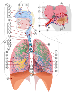
A respiratory system´s function is to allow gas exchange. The space between the alveoli and the capillaries, the anatomy or structure of the exchange system, and the precise physiological uses of the exchanged gases vary depending on the organism. In humans and other mammals, for example, the anatomical features of the respiratory system include airways, lungs, and the respiratory muscles. Molecules of oxygen and carbon dioxide are passively exchanged, by diffusion, between the gaseous external environment and the blood. This exchange process occurs in the alveolar region of the lungs. [1]
Other animals, such as insects, have respiratory systems with very simple anatomical features, and in amphibians even the skin plays a vital role in gas exchange. Plants also have respiratory systems but the directionality of gas exchange can be opposite to that in animals. The respiratory system in plants also includes anatomical features such as holes on the undersides of leaves known as stomata.
Contents |
[edit] Anatomy of respiratory system in Vertebrates
[edit] Mammals
For mammals, including humans, respiration is essential. In these organisms, the respiratory system can be subdivided into an upper respiratory tract and a lower respiratory tract based on anatomical features. The upper respiratory tract includes the nasal passages, pharynx and the larynx, while the lower respiratory tract is comprised of the trachea, the primary bronchi and lungs. The respiratory system can also be divided into physiological, or functional, zones. These include the conducting zone (the region for gas transport from the outside atmosphere to just above the alveoli), the transitional zone, and the respiratory zone (the alveolar region where gas exchange occurs). (See also respiratory tract.)
[edit] Comparative anatomy/physiology in mammals
[edit] Horses
Horses are obligate nasal breathers. That is, they are different from many other mammals in that they do not have the option of breathing through their mouths and must take in air through their nose.
[edit] Elephants
The elephant is the only mammal known to have no pleural space. Rather, the parietal and visceral pleura are both composed of dense connective tissue and joined to each other via loose connective tissue. [2] This lack of a pleural space, along with an unusually thick diaphragm, are thought to be evolutionary adaptations allowing the elephant to remain underwater for long periods of time while breathing through its trunk which emerges as a snorkel. [3]
[edit] Marine Mammals
[edit] Rodents
[edit] Birds
The respiratory system of birds differs significantly from that found in mammals, containing unique anatomical features such as air sacs. The lungs of birds also do not have the capacity to inflate as birds lack a diaphragm and a pleural cavity. Gas exchange in birds occurs between air capillaries and blood capillaries, rather than in alveoli. See Avian respiratory system for a detailed description of these and other features.
[edit] Reptiles
The anatomical structure of the lungs is less complex in reptiles than in mammals, with reptiles lacking the extensive airway tree structure found in mammalian lungs. Gas exchange in reptiles still occurs in alveoli, however. Reptiles do not possess a diaphragm. Thus, breathing occurs via a change in the volume of the body cavity which is controlled by contraction of intercostal muscles in all reptiles except turtles. In turtles, contraction of specific pairs of flank muscles governs inspiration or expiration. [4]
See also reptiles for more detailed descriptions of the respiratory system in these animals.
[edit] Amphibians
The skin is one of the important respiratory organs in amphibians. It is highly vascularized and moist, with moisture maintained via secretion of mucus from specialized cells. These properties aid rapid gas exchange.
[edit] Fish
In most fish the respiration takes place through gills. (See also aquatic respiration.) Lungfish, however, do possess one or two lungs. The labyrinth fishes have developed a special organ that allows them to take advantage of the oxygen of the air, but is not a true lung.The rare breath fish also has two lungs.
[edit] Anatomy of respiratory system in invertebrates
[edit] Sponges and jellyfish
These animals lack specialized organs for gas exchange, instead taking in gases directly from the surrounding water.
[edit] Flatworms and annelids
Flatworms have special muscles, called "enmmustullus", meaning "small muscles" in Latin. These muscles help the worms to create energy efficiently, while still completing essential activities like eating and sleeping.
[edit] Insects
Air enters the respiratory system of most insects through a series of external openings called spiracles. These external openings, which act as muscular valves in some insects, lead to the internal respiratory system, a densely-networked array of tubes called trachea. The tracheal system within an individual is composed of interconnecting transverse and longitudinal tracheae which maintain equivalent pressure throughout the system. These tracheae branch repeatedly, eventually forming tracheoles, which are blind-ended, water-filled compartments only one micrometer in diameter [1]. It is at this level of the tracheoles that oxygen is delivered to the cells for respiration.
Insects were once believed to exchange gases with the environment continuously by the simple diffusion of gases into the tracheal system. More recently, however, large variation in insect ventilatory patterns have been documented and insect respiration appears to be highly variable. Some small insects do demonstrate continuous respiration and may lack muscular control of the spiracles. Others, however, utilize muscular contraction of the abdomen along with coordinated spiracle contraction and relaxation to generate cyclical gas exchange patterns. The most extreme form of these patterns is termed discontinuous gas exchange cycles (DGC) [5].
[edit] Molluscs
Molluscs generally possess gills that allow exchange of oxygen from an aqueous envinronment into the circulatory system. These animals also possess a heart that pumps blood which contains hemocyanin as its oxygen-capturing molecule. Hence, this respiratory system is similar to that of vertebrate fish.
[edit] Physiology of respiratory system in mammals
For more detailed descriptions see also Respiratory physiology or Respiration.
[edit] Ventilation
Ventilation of the lungs is carried out by the muscles of respiration.
[edit] Control
Ventilation occurs under the control of the autonomic nervous system from parts of the brain stem, the medulla oblongata and the pons. This area of the brain forms the respiration regulatory center, a series of interconnected brain cells within the lower and middle brain stem which coordinate respiratory movements. The sections are the pneumotaxic center, the apneustic center, and the dorsal and ventral respiratory groups. This section is especially sensitive during infancy, and the neurons can be destroyed if the infant is dropped and/or shaken violently. The result can be death due to "shaken baby syndrome."[6]
[edit] Inhalation
Inhalation is initiated by the diaphragm and supported by the external intercostal muscles. Normal resting respirations are 10 to 18 breaths per minute, with a time period of 2 seconds. During vigorous inhalation (at rates exceeding 35 breaths per minute), or in approaching respiratory failure, accessory muscles of respiration are recruited for support. These consist of sternocleidomastoid, platysma, and the scalene muscles of the neck.
Under normal conditions, the diaphragm is the primary driver of inhalation. When the diaphragm contracts, the ribcage expands and the contents of the abdomen are moved downward. This results in a larger thoracic volume and negative (suction) pressure (with respect to atmospheric pressure) inside the thorax. As the pressure in the chest falls, air moves into the conducting zone. Here, the air is filtered, warmed, and humidified as it flows to the lungs.
During forced inhalation, as when taking a deep breath, the external intercostal muscles and accessory muscles aid in further expanding the thoracic cavity.
[edit] Exhalation
Exhalation is generally a passive process; however, active or forced exhalation is achieved by the abdominal and the internal intercostal muscles. During this process air is forced or exhaled out.
The lungs have a natural elasticity: as they recoil from the stretch of inhalation, air flows back out until the pressures in the chest and the atmosphere reach equilibrium.[7]
During forced exhalation, as when blowing out a candle, expiratory muscles including the abdominal muscles and internal intercostal muscles, generate abdominal and thoracic pressure, which forces air out of the lungs.
[edit] Circulation
The right side of the heart pumps blood from the right ventricle through the pulmonary semilunar valve into the pulmonary trunk. The trunk branches into right and left pulmonary arteries to the pulmonary blood vessels. The vessels generally accompany the airways and also undergo numerous branchings. Once the gas exchange process is complete in the pulmonary capillaries, blood is returned to the left side of the heart through four pulmonary veins, two from each side. The pulmonary circulation has a very low resistance, due to the short distance within the lungs, compared to the systemic circulation, and for this reason, all the pressures within the pulmonary blood vessels are normally low as compared to the pressure of the systemic circulation loop.
[edit] Gas exchange
The major function of the respiratory system is gas exchange between the external environment and an organism's circulatory system. In humans and mammals, this exchange facilitates oxygenation of the blood with a concomitant removal of carbon dioxide and other gaseous metabolic wastes from the circulation. As gas exchange occurs, the acid-base balance of the body is maintained as part of homeostasis. If proper ventilation is not maintained, two opposing conditions could occur: 1) respiratory acidosis, a life threatening condition, and 2) respiratory alkalosis.
Upon inhalation, gas exchange occurs at the alveoli, the tiny sacs which are the basic functional component of the lungs. The alveolar walls are extremely thin (approx. 0.2 micrometres). These walls are composed of a single layer of epithelial cells (type I and type II epithelial cells) in close proximity to the pulmonary capillaries which are composed of a single layer of endothelial cells. The close proximity of these two cell types allows permeability to gases and, hence, gas exchange.
[edit] Non-respiratory functions
[edit] Vocalization
The movement of gas through the larynx, pharynx and mouth allows humans to speak, or phonate. Vocalization, or singing, in birds occurs via the syrinx, an organ located at the base of the trachea. The vibration of air flowing across the larynx (vocal chords), in humans, and the syrinx, in birds, results in sound. Because of this, gas movement is extremely vital for communication purposes.
[edit] Temperature control
Panting in dogs and some other animals provides a means of controlling body temperature. This physiological response is used as a cooling mechanism.
[edit] Coughing and sneezing
Irritation of nerves within the nasal passages or airways, can induce coughing and sneezing. These responses cause air to be expelled forcefully from the trachea or nose, respectively. In this manner, irritants caught in the mucus which lines the respiratory tract are expelled or moved to the mouth where they can be swallowed.
[edit] Development of respiratory system in animals
[edit] Humans and mammals
The respiratory system lies dormant in the human fetus during pregnancy. At birth, the respiratory system becomes fully functional upon exposure to air, although some lung development and growth continues throughout childhood. Pre-term birth can lead to infants with under-developed lungs. These lungs show incomplete development of the alveolar type II cells, cells that produce surfactant. The lungs of pre-term infants may not function well because the lack of surfactant leads to increased surface tension within the alveoli. Thus, many alveoli collapse such that no gas exchange can occur within some or most regions of an infant's lungs, a condition termed respiratory distress syndrome. Basic scientific experiments, carried out using cells from chicken lungs, support the potential for using steroids as a means of furthering development of type II alveolar cells.[8]In fact, once a pre-mature birth is threatened, every effort is made to delay the birth, and a series of steroid shots is frequently administered to the mother during this delay in an effort to promote lung growth.[9]
[edit] Disease and the respiratory system
Disorders of the respiratory system can be classified into four general areas:
- Obstructive conditions (e.g., emphysema, bronchitis, asthma attacks)
- Restrictive conditions (e.g., fibrosis, sarcoidosis, alveolar damage, pleural effusion)
- Vascular diseases (e.g., pulmonary edema, pulmonary embolism, pulmonary hypertension)
- Infectious, environmental and other "diseases" (e.g., pneumonia, tuberculosis, asbestosis, particulate pollutants): Coughing is of major importance, as it is the body's main method to remove dust, mucus, saliva, and other debris from the lungs. Inability to cough can lead to infection. Deep breathing exercises may help keep finer structures of the lungs clear from particulate matter, etc.
The respiratory tract is constantly exposed to microbes due to the extensive surface area, which is why the respiratory system includes many mechanisms to defend itself and prevent pathogens from entering the body.
Disorders of the respiratory system are usually treated internally by a pulmonologist or respiratory physician1.
[edit] Respiratory system in plants
[edit] Gas exchange in plants
Plants use carbon dioxide gas in the process of photosynthesis, and then exhale oxygen gas, a waste product of photosynthesis. However, plants also sometimes respire as humans do, taking in oxygen and producing carbon dioxide.
Plant respiration is limited by the process of diffusion. Plants take in carbon dioxide through holes on the undersides of their leaves known as stomata (sing:stoma). However, most plants require little air.[citation needed] Most plants have relatively few living cells outside of their surface because air (which is required for metabolic content) can penetrate only skin deep. However, most plants are not involved in highly aerobic activities, and thus have no need of these living cells.
[edit] See also
- Involuntary control of respiration
- Liquid breathing
- Major systems of the human body
- Muscles of respiration
[edit] Notes
- ^ Maton, Anthea; Jean, Hopkins Susan, Johnson Charles William, McLaughlin Maryanna Quon Warner David, LaHart Wright, Jill D. (2009). [= Human Biology and Health]. Englewood Cliffs,: Prentice Hall. pp. 108–118. doi:. =. ISBN 0-12-981176-1. =.
- ^ West, John B.; Ravichandran (1993). "[= Snorkel breathing in the elephant explains the unique anatomy of its pleura]". Respiration Physiology (=: =) 126 (=): 1–8. doi:. =. =.
- ^ West, John B.; = (2002). "[= Why doesn't the elephant have a pleural space?]". News Physiol Sci (=wewe gonzalez: =) 17 (=): 47–50. doi:. =. =.
- ^ Britannica On-line Encyclopedia
- ^ Lighton, JRB; = (January 1996). "[= Discontinuous gas exchange in insects]". Annu Rev Entomology (=: =) 41 (=): 309–324. doi:. =. =.
- ^ *Fact sheet on Shaken Baby Syndrome
- ^ A simple model of how the lungs are inflated can be built from a bell jar
- ^ Department of Environmental Biology, University of Adelaide, Adelaide, South Australia
- ^ Pregnancy-facts.com
[edit] References
- Perkins, M. 2003. Respiration Power Point Presentation. Biology 182 Course Handout. Orange Coast College, Costa Mesa, CA.
- Medical Dictionary
[edit] External links
- Berkeley Anatomy lecture on the respiratory system
- A high school level description of the respiratory system
- Introduction to Respiratory System
- Science aid: Respiratory System A simple guide for high school students
- The Respiratory System University level
|
|||||
|
||||||||
|
||||||||||||||
|
||||||||||||||||||||
|
|||||||||||||||||||||||||||||||||||||||||||||||||||||||||||
|
||||||||

